 Research Article
Abstract
Objective: To evaluate the effectiveness of the Well Wave equipment in treating patients with injuries to the tendons and ligaments of the osteomyoarticular system.
Methods: A descriptive, cross-sectional study was carried out with 107 patients diagnosed with injuries to the tendons and ligaments of the osteomyoarticular system, which were treated with the Well Wave equipment (extracorporeal shock waves) at the International Orthopedic Scientific Complex “Frank Country ”, in the period between March 2019 and April 2020. The analysis of the variables (age, sex, pain and disability) was carried out. To determine the effectiveness (real benefit), the medical and physiological effects that occur when applying this therapy were analyzed.
Results: Female sex predominated (71.9%) and 51-60 years (28%). 40.2% of the patients were treated for presenting supra spinous tendonitis. All patients had pain before the application of therapy. After treatment, 86.9% of the patients stopped feeling it and 58% presented absence of disability according to the DASH scale. According to the evaluation criteria of the response to treatment, the results were satisfactory (72.9%).
Conclusions: The therapy with shock waves enabled a rapid recovery of the patients attended and their incorporation into daily activities.
Keywords: Shock Waves; Supraspinatus Tendinis; Epicondylitis; Heel Spur; Calcific Achillean Tenosynovitis
Introduction
For more than 20 years, medical technology with great potential to improve or replace some invasive procedures has been successfully applied in the world, such is the case of Extracorporeal Shock Wave therapies. In 1997 the European Society for Muscleskeletal Shock Wave Therapy was established in Vienna, due to the rapid diffusion of the method, in 1999 it was renamed the International Society for Muscle-skeletal Shock Wave Therapy [1]. The application of this therapy has proven to be safe and effective, as it has avoided surgical procedures. Shock wave therapy is acoustic waves present in everyday situations (the sound of thunder, clapping in an auditorium, or an airplane breaking the sound barrier). The wave generates a sudden pressure variation that propagates in the three planes of space; it goes from ambient pressure to the maximum pressure peak at the wave front. In the case of its therapeutic application, the wave is transmitted through a coupling pad that is a liquid medium that, having an acoustic consistency similar to that of the human body, favors its transfer. It is essential that there is a transitional medium between the docking pillow and the body, such as ultrasound gel. The shock waves are directed towards a focal point in the tissue to be treated. For the shock wave to have an adequate therapeutic effect, the energy must be focused on the point to be treated. The depth of penetration of the shock wave focus into the tissues can be varied by modifying the thickness of the coupling pillow [1,2]. At present in Cuba the number of elderly people has increased, this brings a series of progressive physiological and functional deteriorations with the consequent acquisition of chronic degenerative diseases. Diseases that cause joint pain are chronic and often disabling. Between 50-80% of the population over 65 years of age present pain [3]. Medical care for osteomyoarticular conditions that end in a surgical procedure is high, every day the number of patients who need specialized medical assistance in search of a solution to their health problem is greater.
Thus, it is necessary to use a new technology that manages to improve, replace or replace some old invasive procedures compared to new technologies, such is the case of Extracorporeal Shock Wave therapies, with the aim of improving the quality of life of patients. the Cuban population and treat conditions of the osteomyoarticular system. In Cuba, at the “Frank País” International Orthopedic Scientific Complex, it began to be applied in 2001. This therapy has been used until now as a method of treating chronic pain in patients who do not improve with other conservative treatments. The use of this equipment in musculoskeletal conditions enables a wave disintegrating effect to treat calcifications, has analgesic effects and allows stimulation of the repair process in tendons, soft tissues and bones [4]. The treatment unit produces, by means of an external piezoelectric source, high-energy sound waves, which have a great depth of penetration. This maximum energy in the affected area allows exact location of the area to be treated, low risk of bruising and very little irritation to the skin. It facilitates the outpatient treatment of the patient, short sessions [3-5], without risk of allergy or the need for anesthesia [5].
The number of patients who have received physiotherapeutic treatment with shock waves for this cause has increased in recent years at our institution. The objective then of our research was to evaluate the effectiveness of extracorporeal shock wave therapy in patients with injuries to the tendons and ligaments of the osteomyoarticular system.
Methods
A descriptive, cross-sectional study was carried out with patients who presented tendon and ligament injuries (supraspinatus tendinis, epicondylar insertions, calcified achillean tenosynovitis, calcaneal spur and plantar fasciitis). For the therapeutic treatment, the Well Wave equipment (extracorporeal shock waves) was used, in the International Orthopedic Scientific Complex “Frank País”, in the period from March 2019 to April 2020. The selection of patients in the sample was carried out through a convenience sampling and was made up of 107 cases. To determine the effectiveness (real benefit), the medical and physiological effects that occur when applying this therapy were analyzed.
Inclusion criteria
a) Patients of both sexes, aged over 18 years.
b) Patients with persistence of pain in a period of six months or more.
c) Patients undergoing failed previous surgery.
d) Patients who received at least three of the following treatments:
e) Medications
f) Infiltrations
g) Laser
h) Therapeutic ultrasound
i) Magnetotherapy
j) Supports
Exclusion criteria
k) Patients who refuse to continue in the study.
l) Patients with decompensated chronic diseases (severe cardiovascular disorders, neurological disorders).
m) Patients with bleeding disorders.
n) Pregnant patients.
o) Patients with polyneuropathies.
p) Patients with epiphysiolysis.
q) Patients with pacemakers.
r) Patients with primary or metastatic malignant tumors.
s) Patients with acute or chronic tissue infections.
t) Patients with severe arthritic changes.
In the initial consultation, all patients underwent a detailed interrogation, physical examination, radiographic study in anteroposterior views and ultrasound of the soft tissues of the affected area. Once the clinical radiological diagnosis was made that they had some injury to the tendons and ligaments (supraspinatus tendinis, epicondylar insertions, calcified achillean tenosynovitis, calcaneal spur and plantar fasciitis) and taking into account that the patient had used other conservative and surgical methods and had not presented improvement, he began with the therapeutic treatment of extracorporeal shock waves in the affected area with the Well Wave equipment, consisting of a piezoelectric shock wave source mounted on a mobile arm with a full range of motion. All patients were asked for their informed consent to participate in the study (Annex 3).
Process
a. Patient lying or sitting in right or left lateral decubitus, depending on location, on a stretcher in the shock wave unit.
b. Localization of the painful point by palpation and lubrication of the treatment area with gel on the skin and on the coupling membrane of the equipment for the transmission of shock waves.
c. Energy density from 0 mj / mm2 to 20 mj / mm2 . Progressive application of shock waves from low intensity to maximum intensity according to tolerance.
d. Frequency 0 - 4
e. Maximum pressure 6 to 126 Mpa.
f. Depth of penetration 0 mm to 165 mm
g. Sessions: 3 to 5 (once a week).
h. Duration of treatment: 20 to 30 min.
i. No sedation or pain relievers
The patients were evaluated by the authors before and after treatment. The following variables were taken into account: age, sex, pain and disability. In addition, specific scales were used for musculoskeletal diseases, supported by studies collected in the scientific literature. For the assessment of pain (VAS analog scale) (Annex 1). The DASH scale (arm, shoulder and hand disabilities) (Annex 2) was used for a global assessment of symptoms and disability; This measures the ability to carry out activities of daily life and work, composed of 30 items, measures different dimensions: functionality, symptoms, social role and psychological state. The score ranges from 0 in those patients in the absence of disability to 100 total disabilities. Its authors proposed a criterion for the qualitative interpretation of the results (Absence: 0, mild disability: 1-10, moderate disability: 11-40, severe disability: 41- 80 and total disability: 81-100) that, despite having received some criticism, it is still used.
The criteria for evaluating response to treatment:
1. Good evolution when there is remission of pain (EVA-0), restoration of joint mobility and incorporation of patients to their usual activities.
2. Regular evolution when there is a conspicuous improvement in pain (EVA ≤3), improvement in joint range of motion, requirement of some conservative treatment and incorporation to their usual activities.
3. Poor outcome when there was no remission of pain (EVA >3), no improvement in mobility and no incorporation to usual activities.
The information processing was carried out in EXCEL and using the statistical package SSPS 11.5. The frequency analysis of the variables contemplated in the study was carried out, the absolute and relative frequencies were obtained. In addition, measures of central tendency (mean, median) and dispersion (standard deviation) were used. Likewise, the Chi square test of independence was performed to determine the existence of statistically significant differences between the proportions obtained. In each case, the value corresponding to the probability of occurrence p; an alpha error of 0.05 and a confidence of 95% were prefixed. It was determined, as a critical or rejection region, when the value associated with p was less than 0.05 and, in this case, the null hypothesis of independence was rejected, and it was concluded that the variables were dependent on each other.
Ethical Considerations
The study was carried out following the ethical principles set out in the Declaration of Helsinki. The Ethics Committee for research in humans, of the International Orthopedic Scientific Complex “Frank País”, ensured compliance with these requirements and approved the research. The information obtained was kept confidential and was only used for investigative purposes. Similarly, to obtain personal information from each patient, their consent was requested, and the reserved nature of the information provided and its scientific use was explained (Annex 3).
Results
As can be seen in Table 1, in the sample there was a predominance of females with 77 patients (71.9%) and the age group of 51-60 years (28%). The median age of the patients seen was 58 years of age. There were no significant differences in terms of age and sex distribution (p = 0.253).
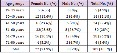
Table 1: Patients with tendon and ligament injury according to age and sex.
p= 0,253
Table 2 shows the distribution of patients with tendon and ligament injury according to sex. 40.2% of the patients were treated for presenting supra spinous tendonitis, followed by patients who were diagnosed with a calcified Achillean Tenosynovitis, 22.4% of the sample. There were no significant differences (p = 0.345).
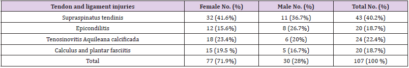
Table 2: Patients with tendon and ligament injury according to sex.
AF: Absolute frequency p = 0.345
As can be seen in Table 3, all the patients had pain before the application of the shock wave, inclusive, it was the main indication for performing this non-invasive technique. After treatment, this situation was reversed, 78.5% of the patients were without pain. Only 8.4% had pain while walking and 7.5% at rest. These differences in pain before and after therapy were statistically significant (p = 0.0000).
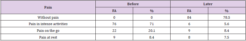
Table 3: Patients with tendon and ligament injury, according to visual analog scale, before and after treatment.
AF: Absolute frequency p = 0.0000
Table 4 shows the results obtained after applying the DASH Scale. 43% of the patients studied had total disability before starting treatment. Only 4.7% were found in the category of absence of disability. After five sessions of therapy, according to the procedure described, 71% had no disability and only 5.6% remained in the category of total disability. Significant results were obtained (p = 0.0000).
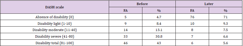
Table 4: Patients with tendon and ligament injury, according to the DASH scale, before and after treatment.
AF: Absolute frequency p = 0.0000
Table 5 shows the analysis of the evaluation criteria for response to treatment, where good and fair results were considered satisfactory and bad, unsatisfactory. There was a higher percentage of satisfactory results (56% and 23.4%). Only 20.6% of the sample had a poor response, persisting symptoms of pain and functional limitation. The results were significant (p = 0.0000).
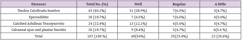
Table 5: Evaluation criteria of the response to the treatment given to the patients.
Discussion
According to the literature reviewed, tendinopathies of the shoulder injury to the supraspinatus, tennis elbow (epicondylitis) are common between 40 and 60 years of age. The literature states that 2 to 50% of the population have shoulder pain, accompanied by common symptoms such as atrophy of the muscles and limitation of movements. It is common in both sexes, with a 4: 1 ratio, in favor of women associated with jobs such as seamstresses, housewives, hard workers, athletes and musicians. In the case of Epicondylitis, its prevalence is 10% [6]. Calcaneal spur, plantar fasciitis and calcific achillean tenosynovitis are diseases that have a high prevalence that increases with age. It is of multifactorial origin, although a history of repetitive microtraumas is collected, being more common in runners, overweight people and tasks that require standing for long periods of time. It affects 10% of the population throughout their life between the fourth and sixth decade of life, also causing functional disability. The aforementioned diseases are common conditions in women, which coincided with our study, including age [7]. Age can be considered a risk factor in itself for the suffering of these conditions, since in the aging process itself changes occur in our body that favor the appearance of these diseases.
According to Mirallas Martínez, pain, limited movement and disability is a frequent symptom, characteristic of all these conditions and can be present in all cases [8]. This result could be corroborated in our investigation. The pain can be present, even in a state of rest, and it can make it impossible to sleep at night if you sleep on the affected side. Night pain can be severe enough to prevent sleep or wake the patient at night. Extracorporeal shock waves pass through tissues and can trigger absorption, reflection, refraction and energy transmission phenomena (direct effect). The negative phase is the cause of the indirect effects on the cellular tissue. These types of waves increase metabolism in the body and favor the reduction of inflammation in the area affected by the production of endorphins, causing a triggering analgesic action. In this way, the process of stimulation of inflammation mediators by induced hyperemia and the release of free radicals is accelerated.
The reversal of chronic inflammation is another advantage of the use of extracorporeal shock waves, since this persistent inflammation requires components (called mast cells), whose activity increases with acoustic waves, allowing the production of chemokines and cytokines that improve the inflammatory process [9]. The DASH questionnaire was developed in 1994 in English at the initiative of the American Academy of Orthopedic Surgeons (AAOS), as a consequence of this, from 1994 to the present, more than 48 validated and culturally adapted versions of the DASH have been developed (website of Institute for Work & Healt: http://www. dash.iwh.on.ca) is specific for musculoskeletal conditions. Although it is an instrument used to evaluate only disability in the upper limbs, it was of great value to the authors, since a greater number of patients with these conditions were diagnosed in the sample, which was essential to use. In addition, this specific instrument made it possible to detect the clinical changes of interest in the patients’ status, it allowed to functionally assess the joint of the shoulder, elbow, wrist and fingers and the quality of life of the patients [10].
Indications for shock wave therapy encompass a wide range of conditions, including (Supraspinatus tendinis, Epicondylar insertions, Calcific Achillean Tenosynovitis, Calcaneal spur, and Plantar fasciitis), among others. Its effectiveness lies in the physiological effects that occur in the body when applying this wave. It causes a regulation of the inflammatory cascade where it decreases the levels of substance P, bradycin and other local factors of inflammation. In addition, it stimulates the phenomenon of angiogenesis, all these changes finally activate and modulate the healing cascade, thus turning a chronic wound into an acute wound that will have a normal physiological healing process [11]. Wang CJ and others in their clinical studies have shown increased blood flow and growth factors that induce angiogenesis and consequent neovascularization in the calcaneal tendinous insertion [12]. It was manifested in the clinical improvement of the patients after applying it, which coincided with our investigation.
Effectiveness was demonstrated in the treatment of supraspinatus tendonitis and epicondylitis [13]. The improvement is significant in pain intensity, with good or excellent functional gain in 56% of patients treated with extracorporeal shock wave therapy. There are significant differences between patients in the treatment and placebo groups in pain and function, and it is concluded that treatment using this therapy is a pre-surgical alternative. The improvement in pain and function is good or excellent in 48% and acceptable in 42%, with a significant difference in favor of patients in the treated group compared to those in the placebo group. The improvement in pain and function is good or excellent in 52% of those treated compared to 6% of those in the placebo group [14]. Similar results to ours were observed.
In the case of plantar fasciitis and achillean tenosynovitis, Gollwitzer H, et al. [15] Conducted the study that used the largest sample size, with 250 subjects for the trial obtaining satisfactory results. This effectiveness or benefits are reflected in an improvement in the patient’s symptoms, especially in the decrease in pain and improvement in functional capacity, and in a decrease in the thickness of the plantar fascia based on the studies reviewed, as these are the variables more measures together [15]. The main weakness of this study is the small sample size, but with results similar to those found in other studies where a similar response to both treatments could be observed, this fact supports the strength of our findings.
Conclusion
Some of the sociodemographic characteristics of the patients studied do not differ much from those indicated by other authors, such as: the predominance of female sex and age. Treatment with extracorporeal lithotripsy (shock wave therapy), with the Well Wave equipment, was an effective method. It is a modern and noninvasive technique, which has enabled a rapid recovery of patients, their incorporation into daily activities, promotes rehabilitation, early return to work activities and better use of the working day.
References
- Schmitz C, Császár NBM, Milz S, Schieker M, Maffulli N, et al. (2015) Efficacy and safety of extracorporeal shock wave therapy for orthopedic conditions: A systematic review on studies listed in the Pedro database. Br Med Bull 116(1): 115-138.
- Wang CJ (2012) Extracorporeal shockwave therapy in musculoskeletal disorders. J Orthop Surg Res 7: 11-8.
- Carrington Reid M, Eccleston Ch, Pillemer K (2015) Management of chronic pain in older adults. BMJ 350: h532.
- García Estrada E, Álvarez Cambras R, Rodríguez Vázquez M, Valdés Díaz A, González Fundora N (2005) Plantar fasciitis treated with extracorporeal shock waves. (Fascitis plantar tratada con ondas de choque extracorpóreas). Rev Cubana Ortop Traumatol 19(1).
- Español Barrull AR (2014) Extracorporeal shock waves in the treatment of nonunions of the long bones of the lower extremities. (Ondas de choque extracorpóreas en el tratamiento de las pseudoartrosis de los huesos largos de las extremidades inferiors). [Tesis de grado]. Barcelona, España: Universitat Internacional de Catalunya).
- Garcés J (2016) Efficacy of shock wave therapy as a treatment alternative for rotator cuff injuries. Duazary. (Eficacia de la terapia de ondas de choque como alternativa de tratamiento en lesiones del manguito rotador. Duazary) 13(1): 23-29.
- Lim AT, How CH, Tan B (2016) Management of plantar fasciitis in the outpatient setting. Singapore Medical Journal 57(4): 168-171.
- Mirallas Martínez JA (2005) Evidence-Based Extracorporeal Shock Wave Effectiveness. (Efectividad de las ondas de choque extracorpóreas basada en la evidencia). Rehabilitación (Madr). 39(2): 52-58.
- Romero M Martínez A (2015) Scope of extracorporeal shock wave therapy in musculoskeletal injuries. Venezuelan Archives of Pharmacology and Therapeutics (Alcances de la terapia con ondas de choque extracorpóreas en lesiones músculo-esqué Archivos Venezolanos de Farmacología y Terapéutica) 34(2): 27-30.
- Tongprasert S, Rapipong J, Buntragulpoontawee M (2015) The cross – cultural adaptation of the DASH questionnaire in Thai (DASH –TH). J Hand Ther 27(1): 49-54.
- Martín Cordero J (2008) Therapeutic Physical Agents. Havana: Medical Sciences Editorial (Agentes Físicos Terapé La Habana: Editorial Ciencias Médicas) 23(1): 246-251.
- Wang CJ, Huang HY, Pai CH (2002) Shock wave-enhanced neovascularization at the tendon-bone junction: an experiment in dogs. The Journal of foot and ankle surgery: official publication of the American College of Foot and Ankle Surgeons 41(1): 16-22.
- Haist J (2000) Shockwave treatment for radial and ulnar Epicondylitis. Musculoskeletal Shockwave Therapy. London: Greenwich Medical Media p.110-114.
- Haake M, Deike B, Schmitt J (2002) Exact focusing of extracorporeal shockwave therapy for calcifying tendinopathy. Clin Orthop (397): 323-331.
- Gollwitzer H, Saxena A, DiDomenico LA, Galli L, Bouche RT, et al. (2015) Clinically relevant effectiveness of focused extracorporeal shock wave therapy in the treatment of chronic plantar fasciitis: a randomized, controlled multicenter study. The Journal of bone and joint surgery American volume 97(9): 701-708.
