 Research Article
Abstract
Angiogenesis is the formation of new blood vessels from main blood vessel. The chorioallantoic membrane model has been frequently used method for functionally beneficial aspects of demonstration of the angiogenesis and understanding of the cancer pathogenesis. Acesulfame-K is an artificial sweetener and about 120 times sweeter than sucrose. It is not metabolized in the human body, so it does not provide calories. Saccharin is also an artificial sweetener and about 300 times sweeter than sucrose. Some studies have shown a relationship between saccharin consumption and cancer incidence. Therefore, we aimed to investigate the effect of acesulfame-K and saccharin on angiogenesis by using in vivo CAM assay.
Keywords: Sweeteners; Acesulfame-K; Saccharin; Angiogenesis; Chorioallantoic Membrane Assay
Introduction
Angiogenesis is defined as the formation of new blood vessels due to growth, injury, hypoxia, or nutritional stress in a pre-existing vasculature [1,2]. In addition, the angiogenic process is responsible for many pathologic processes including diabetic retinopathy, haemangiomas, solid tumour growth and fibrosis [3,4]. It is a fact that angiogenesis is the equilibrium between angiogenic substances that create new vessels and antiangiogenic substances that inhibit new vessel formation factors [5]. The CAM model is a simple, rapid and cost-effective in vivo model that does not require ethics committee approval, which allows monitoring tumor growth and metastasis, wound healing, drug distribution, and angiogenic and antiangiogenic molecules [6,7]. Acesulfame-K is an artificial sweetener and about 120 times sweeter than sucrose. It is not metabolized in the human body, so it does not provide calories. A disintegration product of Ase-K is Acetoacetamide, which is toxic when consumed at very high doses [8]. Acesulfame-K’s acute toxic effect on humans is headache, while chronic toxic effects are clastogenic and genotoxic in high doses [9]. Saccharin is also an artificial sweetener and about 300 times sweeter than sucrose. The FDA attempted to ban saccharin in 1977, when animal studies showed that it caused cancer in mice (mostly bladder cancer). Since then, many studies have been carried out on saccharin. No study showed a clear association between human saccharin consumption at normal doses and health risks. Some studies have shown a relationship between consumption and cancer incidence [8,9].
Material and Method
Material
Egg Supply: ATAK-S type fertilized chicken eggs were taken from Istanbul chicken farm. All egg surfaces were cleaned with ethyl alcohol due to contamination risk. We kept the eggs under optimal conditions (60% relative humidity at 37.5°C) in an incubator.
Sweeteners: The active compounds, acesulfame-K (lot: 1610017) and Saccharin sodium (lot: 15041802) were obtained from Prinova Food and Chemical Trade Limited Company. Acesulfame-K (MA: 201.2) 1,006 g and Saccharin sodium (MA: 205.17) 1.025 g were weighed into solutions of 100 ml 0.9% Isotonic Saline at concentrations of 50 mM.
Method
Chorioallantoic Membrane Assay (CAM): Chorioallantoic Membrane Assay is an easy way to observe changes on angiogenesis, results fast and inexpensive. Therefore, in this study, we used Chorioallantaoic Membrane (CAM) model in chick embryos. We divided eggs into three groups as A, B and C and put them inside of an incubator. On the 5th day of incubation, the upper part of the eggshells removed without damaging or contaminating the embryo with help of a flashlight. Then we covered removed sites with parafilm due to contamination risk and put them back into incubator to keep temperature optimal for the embryos.
Applying Acesulfame-K and Saccharin Sodium to the Eggs: Acesulfame-K applied to group A eggs, Saccharin sodium applied to group B eggs, and nothing applied to group C for being our control group. Right after application, we parafilmed eggs again and returned back to the incubator for another 2 days. On the 5th day (application time of Acesulfame-K and Saccharin sodium), we took photos, then on the 7th day (after 2 days of application of Acesulfame-K and Saccharin sodium), we removed parafilm from the eggs to observe changes on vascular structure and took photos them for evaluating the results.
Results
72 eggs were used in this study. During the study, 15 eggs were noticed death. On the 5th day, Acesulfame-K solution was applied to 27 eggs in group A. Saccharin sodium solution was applied to 28 eggs in group B. 2 eggs were divided into control group (group C). On the 2nd day after the application of sweeteners, eggs were evaluated macroscopical by 4 observers. Eggs were scored with Knighton et al.’s scoring methodology [10]. Sweeteners caused a significant increase in the blood vessels of the CAMs compared to the control group (Figures 1-3). Changes on angiogenesis defined quantitatively shown in Table 1. The macroscopic evaluation results of CAMs of eggs were shown in Table 2.
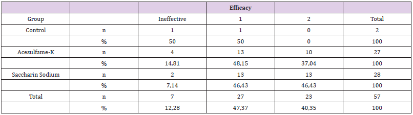
Table 1: After the observation macroscopically, changes on angiogenesis defined quantitatively.
Table 2: Macroscopic evaluation of the effect of sweeteners on CAM.
Note: CAM: Chorioallantoic Membrane
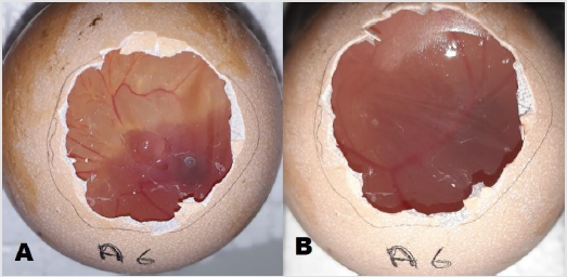
Figure 1: A. Before injection of Asesulfam K. B. After injection of Asesulfam K. As seen macroscopically angiogenesis increased (+1) after injection.
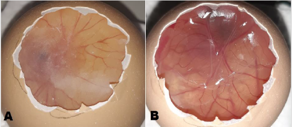
Figure 2:A. Before injection of Sodium Saccharin. B. After injection of Sodium Saccharin. As seen macroscopically angiogenesis increased significantly (+2) after injection.
Figure 3:There is no any injection to A and B. As seen macroscopically there is no significant changes of angiogenesis status.
Discussion
The chorioallantoic membrane model has been frequently used method for functionally beneficial aspects of demonstration of the angiogenesis and understanding of the cancer pathogenesis. Since literature on the subject lack’s studies that research about the effects of specific artificial sweeteners on the angiogenesis, this study holds very important place to understand the relationship between each artificial sweeteners, angiogenesis and also cancer. Aspartame is the one of the very well-known artificial sweeteners that effects on angiogenesis and cancer pathogenesis is studied mostly. One of these studies Yesildal et al. conducted a study in two different methods to see the effects of aspartame on angiogenesis according to colorectal cancer. They used both in vitro model and in vivo CAM model which is a same model used in our study. In addition, results of both studies are same, and study concluded that aspartame has pro-angiogenic effect which is a similar result with our study [11]. In another research, Alleva et al. studied the effect of aspartame which is an artificial sweetener, in angiogenesis on in vitro model and showed that aspartame induced regenerative cytokine production leading to the activation of MAPKs and resulting in the angiogenesis [12]. The difference between our study and this study lay on the method of the studies. We used in vivo CAM model for the examination of the angiogenesis and tried 2 different artificial sweeteners to see both effects compared to the control group.
However, our study differentiates from other artificial sweeteners on angiogenesis studies with the usage of different types of artificial sweeteners. We used Acesulfame-K and Saccharin Sodium which effects on angiogenesis are not very well known. Our study showed that artificial sweeteners like Acesulfame-K and Saccharin Sodium have potential positive effects on angiogenesis. In contrast to our study results, Dong et al. studied the effect of dietary intake of sugar substitutes on endothelial progenitor cells during a cerebrovascular attack and concluded that long-term consumption of sugar substitutes can be responsible of the aggravation of the cerebral ischemic injury. Also, he stated that this effect of sweeteners can be caused by the reduction of the angiogenesis [13]. However, the sweeteners used in this study are generally different from sweeteners that used in our study. Only acesulfame K was the common agent used in both studies and the difference between the results can be caused by the research’s examinations associated with the cerebral ischemic injury. Our study didn’t make any associations with any disease, and it shows the direct effects of the artificial sweeteners on angiogenesis on CAM model. However more studies are needed to understand the effects of Acesulfame-K and Saccharin Sodium on angiogenesis.
Conflict of Interest
None.
References
- Vedat Yildirim, Suat Doganci, Fatih Yesildal, Erkan Kaya, Mehmet Emin Ince (2015) Sodium Nitrite Provides Angiogenic and Proliferative Effects In vivo and In vitro.
- Adam C Mirando, Khadar Abdi, Peibin Wo, Karen M Lounsbury (2017) Assessing the effects of threonyl-tRNA synthetase on angiogenesis-related responses.
- Ataergin AS, Ozet A, Arpacı F (1999) The place of angiogenesis inhibitors in cancer therapy. Turk Klin J Med Sci 19: 100-105.
- Samet C, Musa E, Ebrar B, Elif A, Muaz E, et al. (2020) Effects of Gold Nanoparticles on Angiogenesis in A Chick Chorioallantoic Membrane Model. 10.14235/bas.galenos.2020.3668. Bezmialem Science 8(4): 398-402.
- M Moehler , AD Ho, H Goldschmidt, B Barlogie (2003) Angiogenesis in hematologic malignancies. Crit Rev Oncol Hematol 45(3): 227-244.
- Domenico Ribatti, Roberto Tamma (2018) The chick embryo chorioallantoic membrane as an in vivo experimental model to study human neuroblastoma.
- Domenico Ribatti (2016) The chick embryo chorioallantoic membrane (CAM). A multifaceted experimental model.
- Chattopadhyay S, Raychaudhuri U, Chakraborty R (2014) Artificial sweeteners–a review. Journal of food science and technology 51(4): 611-621.
- Whitehouse CR, Boullata J, McCauley LA (2008) The potential toxicity of artificial sweeteners. Aaohn Journal 56(6): 251-261.
- Knighton D, Ausprunk D, Tapper D, J Folkman (1977) Avascular and vascular phases of tumour growth in the chick embryo. Br J Cancer 35(3): 347-356.
- Yesildal F, Aydin FN, Deveci S, Tekin S, Aydin I, et al. (2015) Aspartame induces angiogenesis in vitro and in vivo models. Hum Exp Toxicol 34(3): 260-265.
- Alleva R, Borghi B, Santarelli L, Strafella E, Carbonari D, et al. (2011) In vitro effect of aspartame in angiogenesis induction. Toxicol In Vitro 25(1): 286-293.
- Dong XH, Sun X, Jiang GJ, Chen AF, Xie HH (2015) Dietary intake of sugar substitutes aggravates cerebral ischemic injury and impairs endothelial progenitor cells in mice. Stroke 46(6): 1714-1718.
