 Research Article
ABSTRACT
The main purpose of this study was to observe the effect of Solanine and Curdione on HIF-1α in renal carcinoma transplanted tumor and human renal carcinoma ACHN cells, and to detect the upstream regulatory proteins of HIF-1α (PI3K/p-PI3K, Akt/p- Akt, mTOR), and to explore the anti-renal cancer mechanism of Solanine and Curdione. Research is divided into two parts, the first part of the research in kidney transplantation tumor, the method is: take the subcutaneous injection method to establish nude mouse model of kidney transplantation tumor ACHN cells, divided into the Solanine, Curdione, Solanine&Curdione (1:1) group and the negative control group, the abdominal cavity injection drug delivery 1 times a day, for 28 days to death mice, tumors present organization, said the tumors had the quality, calculate the inhibitory rate. The expression of HIF-1α protein was detected by Western blotting. The content of ATP in tumor tissues was determined by biochemistry method. The expressions of p-PI3K, p-Akt, mTOR and other proteins were detected by Western Blot. The results showed that all nude mice were successfully molded, and no nude mice died during the medication period.
Compared with the control group, Solanine, Curdione and combination groups significantly decreased the expression of HIF-1α protein in tumor tissues (P < 0.05), and the combination group had a more significant down-regulation effect (P < 0.01). Both Solanine and Curdione could inhibit tumor growth in tumor-bearing nude mice (P < 0.05), and the combination of Solanine and Curdione had a more significant antitumor effect (P < 0.01). Solanine and Curdione could decrease the ATP content in tumor tissues (P < 0.05), and the ATP content in combination group was more significantly decreased than that in single drug group (P < 0.01). The second part was the study of renal cancer cells. The specific methods were as follows: cultured human renal cancer ACHN cell lines, treated with Solanine, Curdione, Solanine&Curdione (1:1) and normal saline, respectively. Western blotting method was used to detect the expression of HIF- 1α protein, CCK-8 method was used to determine the inhibition rate of cell proliferation, and the content of intracellular ATP was determined by ELISA method.
The results showed that the relative expression rate of HIF-1α protein in ACHN cells treated with Solanine and Curdione decreased compared with the control group (P < 0.05), and the combined administration of Solanine and Curdione had a more significant down-regulation effect on HIF-1α protein (P < 0.01). Solanine and Curdione significantly inhibited the proliferation of ACHN cells in vitro (P < 0.05), and the combination of Solanine and Curdione significantly inhibited the growth of ACHN cells (P < 0.01). The ATP production of ACHN cells after treatment with Solanine and Curdione was significantly decreased (P < 0.05), and the effect of combined treatment group was more significant than that of single treatment group (P < 0.01). The relative expression rates of p-PI3K, p-Akt and mTOR proteins in ACHN cells treated with Solanine and Curdione were lower than those in the control group (P < 0.05), and the combined administration had a significant down-regulation effect on p-PI3K, p-Akt and mTOR proteins (P < 0.01). Based on the above, we believe that the combination of Solanine and Curdione can significantly inhibit the energy metabolism of renal cancer, mainly by significantly down-regulating the expression of HIF-1α and inhibiting the PI3K/p-PI3K and Akt/p-Akt transformation process of the signal transduction pathway upstream of HIF-1α.
Keywords: Solanine; Curdione; Kidney Cancer; HIF - 1 Alpha; Role; Mechanism
Introduction
Hypoxia-inducible factor-1 (HIF-1) is a protein complex composed of two similar subunits, HIF-1α and HIF-1β. HIF-1 has biological roles including mediating hypoxia and regulating metabolism. HIF-1α is a hypoxia-induced transcription factor that is present at high levels in malignant solid tumors, but not in normal tissues or slow-growing tumors. In fast-growing tumors, HIF-1α is involved in the activation of a number of cellular processes, including resistance to apoptosis, overexpression of the drug envelope pump, vascular remodeling and angiogenesis, and metastasis. In malignant tumor cells, HIF-1α induces overexpression and enhanced activity of several glycolytic protein subtypes. The enhancement of tumor glycolytic flux induced by HIF-1α is also involved in the kinetic changes of key glycolytic enzyme expression isomers. Solanine and Curdione are the main components of the plant Solanine and Curcuma, which have definite anti-malignant effects. Studies have shown that they can significantly inhibit the growth of a variety of malignant tumors. Previous studies have found that Solanine and Curdione can inhibit the growth of kidney energy metabolism process of interference with, but the specific mechanism is unclear, thus, to explore the Solanine, Curdione, the function of kidney HIF - 1 alpha and further study of related mechanisms, for the development and application of Solanine, Curdione has important value, and Solanine, Curdione will be safe and effective prevention and control of malignant tumor of cheap drugs [1-3].
Materials and Methods
Main Materials and Instruments
Human renal carcinoma ACHN cell line was preserved by the Central Laboratory of Shanxi Integrated Traditional Chinese and Western Medicine Hospital. 32 male BALB /c nude mice (SPF grade) aged from 5 to 6 weeks were purchased from Beijing Vitong Lihua Experimental Animal Technology Co., Ltd. Certificate No. SCXK (Beijing) 2018-0004. Shanghai Yuanye Biotechnology Co., Ltd., the purity is greater than 99%. DMEM medium and calf serum were purchased from Hyclone Corporation, USA. CCK8 kit was purchased from Dongren Chemical Technology Co., Ltd. ATP content test box was purchased from Nanjing Jiancheng Institute of Biological Engineering. Actin antibody and hypoxia-inducible factor-1α antibody are products of Cell Signaling Inc [4-6].
Main Methods
In Vivo Experiment
Preparation of Xenograft Tumor Model of Human Renal Carcinoma in Nude Mice: A total of 32 BALB/c nude mice aged 5 to 6 weeks and weighing 18 to 20 g were selected. ACHN human renal carcinoma cells in logarithmic growth phase were adjusted with RPMI 1640 medium into 5×10⁷ single-cell suspension. 0.2 mL cell suspension was subcutaneously inoculated in the left anterior axilla of BALB/c nude mice to establish the xenograft tumor model of ACHN human renal carcinoma nude mice.
Grouping and Dosing Method: 7 days after inoculation ACHN cells (at least the vaccination site subcutaneous tumor nodules can hit), all the animals were randomly divided into negative control group (normal saline), Solanine group (20 mg/Kg), Curdione group (20 mg/Kg), combined treatment group (20 mg/Kg + Solanine Curdione 20 mg/Kg), intraperitoneal injection of 8 animals in each group are to give medicine (10 mL/Kg), 1 times a day, for 28 consecutive days.
Observation of Tumor Volume and Tumor Inhibition Rate: At the end of administration, mice were killed, tumor mass was completely stripped, tumor mass was weighed, and tumor inhibition rate was calculated according to the following formula: tumor inhibition rate (%) = (1- average tumor weight in the treatment group/average tumor weight in the control group) ×100%. ATP Level Detection in Tumor Mass Tissue: The transplanted tumor was taken, weighed and prepared into 10% homogenate with normal saline, and the ATP content in the transplanted tumor tissue was detected according to the kit instructions.
Alpha HIF - 1 Protein Expression in Tumor Tissue of Detection: transplanted tumor, weighing 100 g after extraction of proteins, lines of sds-page electrophoresis (polyacrylamide gel electrophoresis), to set the conditions for 80 V, 25 min, 100 V, 80 min, the end will be transferred to the PVDF membrane protein, set conditions to 200 mA, 180 min, and in TBST containing 5% skimmed milk powder, enclosed 1 h at room temperature. Primary antibody HIF-1α (1∶1 000) and β-actin (1∶5 000) were incubated at 4 ℃ overnight. Wash 3 times with TBST. The second antibody was incubated at room temperature for 2 h. Wash 3 times with TBST.
In Vitro Experiment
Cell Culture: ACHN cells were cultured in DMEM containing 10% calf serum, 100 units /mL penicillin and 100μg/mL streptomycin. The cells were incubated at 37℃ and 5%CO2. The cells were replaced every 2 ~ 3 days and passed every 5 days [7- 10].
CCK-8 Assay was used to Detect the Inhibition Rate of Cell Proliferation: ACHN cells were digested with 0.25% trypsin and suspended in DMEM medium containing 10% calf serum to prepare single-cell suspension with cell density of 5×10⁴/ mL. The cells were segregated into 96-well plates at 100μL per well and incubated in an incubator at 37℃ and 5%CO2 for 24h. Solanine (4μg/mL), Curdione (50μg/mL), Solanine+Curdione (4μg/mL) and Solanine+Curdione (50μg/mL) were added to each well, and a blank control group was set without drugs. Six duplicate Wells were set in parallel for each group. CCK-8 experiment was conducted after 24, 48 and 72h culture. Add 10μl CCK-8 solution to each well and continue to culture at 37℃ and 5%CO2 for 2 h. The absorbance value (OD value) at 450 nm was measured by automatic microplate analyzer. The inhibition rate of cell proliferation (IR) was calculated according to the following formula: IR= (1- mean OD value of the experimental group/mean OD value of the control group) ×100%. Western Blot Analysis of Hypoxia Inducible Factor 1α (HIF- 1α) Protein Levels: Collect groups of cells, to extract protein, according to the results of the protein quantitative packing protein, protein will repackaging good put in boiling water to boil modified 5 min, and electrophoresis separation, enrichment, protein, protein electrophoresis to separate, transferred to the PVDF membrane, with 5% skimmed milk powder, closed in 37 ℃ oven for 2 h, diluent dilution Ⅰ resistance using antibody, 4 ℃ for the night, incubation TTBS membrane washing 6 times every time (5 min), using antibodies diluent dilution tag Ⅱ resistance, incubation in 37 ℃ oven for 2 h, TTBS wash three times, wash 1 TBS, chemiluminescence mixture, Alpha Innotech system was used to scan the PVDF membrane and analyze the image.
ATP Content Determination: Before cell culture and grouping, 48h after administration, the supernatant was discarded by centrifugation, and the cell suspension was made with normal saline, and then the double steamed water was added. After homogenate was broken in the boiling water bath and heated for 10min, the supernatant was extracted by centrifugation for determination. See ATP Kit Instructions for detailed procedures.
The Expressions of p-PI3K, p-Akt, mTOR and Other Proteins were Detected by Western Blot: The supernatant was discarded after ACHN cells adhered to the wall, and all cells were collected after 24h incubation with dosing respectively. The cells were washed twice with PBS at 4℃. The pre-cooled cell lysis buffer was added, and the lysis was carried out on ice for 15min. The lysates were centrifuged at 10000×g at 4℃ for 30min. The protein concentration of supernatant was measured by Bradford method. The protein was separated by 15% SDS-polyacrylamide gel electrophoresis. After electrophoresis will be transferred to nitrocellulose membrane protein, will use 5% skimmed milk powder solution 37 ℃ close after 2 h, respectively with p - PI3K, p - Akt, mTOR a react to 2 h, 37 ℃ PBST clean 2 times, each time 15 min, then 37 ℃ with horseradish enzyme labeled second antibody response 1 h, after washing, fully with PBST to DAB chromogenic, scan, use of actin antibody consistency on the sample amount is determined.
Statistical Treatment
SPSS20.0 was used for statistical analysis, and the experimental data were represented as the experimental data. Analysis of variance was used for comparison between multiple groups, and P <0.05 was considered statistically significant.
Results
In Vivo Experiment
Effect on the Growth of Transplanted Tumor in Nude Mice with Tumor Bearing: After 28 days of treatment with Solanine and/or Curdione, the tumor weight was significantly lower than that of the control group (P<0.01), and the combination group was lower than that of Solanine and/or Curdione alone (P<0.05), as shown in Table 1.
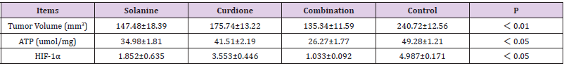
Table 1: Effects of Solanine and/or Curdione alone and in combination on renal carcinoma xenograft tumor related indicators.
Effect of ATP on Tumor Tissue in Nude Mice with Tumor: After 28 days of treatment with Solanine or Curdione, ATP levels in the transplanted tumor tissues decreased, with statistical difference compared with the blank control group (P < 0.05), and the combined group was lower than the single drug group, as shown in Table 1.
Effect of HIF-1α Protein Expression in Tumor Tissues of Nude Mice: Western blotting was used to detect the changes of HIF-1α protein expression in tumor block tissues of each group. The results showed that the HIF-1α protein expression in tumor block tissues of the Solanine and or Curdione intervention group was significantly decreased compared with the blank control group (P < 0.05), and the decrease in the combination group was more significant than that in the single drug intervention group (P < 0.05), Table 1.
In Vivo Experiment
Effects on the Proliferation of ACHN Cells: After treatment with Solanine and Curdione for 24, 48 and 72h, CCK-8 kit was used to detect ACHN cells, and the results showed that the cell growth was inhibited to varying degrees in a time-dependent manner (P < 0.01), as shown in Table 2.
Effect on HIF-1α Protein Expression in ACHN Cells: Western blot results showed that the relative expression rate of HIF-1α protein in ACHN cells treated with Solanine and Curdione was significantly lower than that in the control group (P < 0.05), and the combined administration of Solanine and Curdione significantly down-regulated HIF-1α protein (P < 0.01), as shown in Table 2.
Effects of Solanine and Curdione on ATP Production in ACHN Cells: Results As shown in Table 2, after treatment with Solanine and Curdione, ATP production of ACHN cells decreased (P < 0.05), and the effect of the combined treatment group was more obvious than that of the single treatment group (P < 0.01).
Effects of Solanine and Curdione on p-PI3K, p-Akt and mTOR Protein Expression Levels in ACHN Cells: Western blot results were shown in Table 2. The relative expression rates of p-PI3K, p-Akt and mTOR proteins in ACHN cells treated with Solanine and Curdione were significantly lower than those in the control group (P < 0.05), and the combined administration had a significant down-regulation effect on p-PI3K, p-Akt and mTOR proteins (P < 0.01).
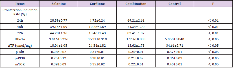
Table 2: Effects of Solanine and/or Curdione alone and combined intervention on ACHN cell related indicators of renal cancer.
Discussion
Hypoxia-inducible factor 1α (HIF-1α) is a type of transcription regulator widely found in humans and mammals under hypoxic environment. It plays an important role in hypoxia-related physiological and pathological processes by activating the transcription activity and expression of different target genes. HIF- 1α is directly or indirectly involved in many processes related to tumor growth, such as tumor proliferation, apoptosis, invasion and metastasis, tumor angiogenesis and tumor drug resistance. The proliferation and metastasis of malignant tumors are rapid and uncontrolled, with insufficient blood supply, insufficient oxygen penetration to meet metabolic requirements, and severe hypoxia in local tissues. At this time, HIF-1α in tumor cells shows the effect of high expression. HIF - 1 alpha to participate in the glucose metabolism of tumor cells, tumor cell metabolism ability is higher than the surrounding normal tissues and its metabolic characteristic is by glycolysis of glucose to their own needs a lot of energy, tumor growth is the lack of blood supply area rely on glycolytic energy, and the way of HIF - 1 alpha is mainly responsible for regulating the expression of glucose transporter protein and hexokinase [11-14].
HIF-1α is highly expressed in tumor cells, which can up-regulate 1/3 expression of glucose transporter and increase glucose uptake. At the same time, HIF-1α can up-regulate the expression of most enzymes in glycolysis and can activate several steps of glycolysis. For example, HIF-1α can upregulate pyruvate dehydrogenase kinase 1 and inhibit pyruvate dehydrogenase, thus inhibiting the tricarboxylic acid cycle, i.e., inhibiting the oxidative phosphorylation pathway. HIF is active in hypoxic tumor cells, and the regulation of enzymes runs through the whole process of glycolytic metabolism. Most of the key enzymes in glycolytic metabolism are positively regulated by HIF-1α. HIF-1α can also regulate the transcription of a variety of target genes and affect the metabolism, proliferation and apoptosis of tumor cells, so that tumor cells can better adapt to the hypoxia microenvironment, and promote tumor angiogenesis, increase the ability of invasion and metastasis, and improve the role of resistance to radiotherapy and chemotherapy.
Solanine and Curdione are the main medicinal ingredients of the plant Solanine and Curcuma, which are also found in other plants. They have definite anti-tumor effects. Studies have shown that they have obvious inhibitory effects on the growth of various tumors. The results of this study showed that Solanine and Curdione could inhibit the proliferation of human renal carcinoma ACHN cells in vitro, reduce the expression of HIF-1α protein, and reduce the production of ATP. Similarly, Solanine and Curdione significantly inhibited the growth of ACHN xenograft tumor in human renal carcinoma in vivo, showing good anti-tumor effects in vivo, significantly reduced the expression of HIF-1α protein in renal carcinoma xenograft tissue, and reduced the generation of ATP in xenograft tumor. Therefore, we proposed the hypothesis that Solanine and Curdione can inhibit tumor metabolism by inhibiting HIF-1α to reduce energy metabolism of tumor cells. It has been reported that HIF-1α synthesis is mediated by activation of the PI3K pathway. The PI3K/ Akt pathway activates mammalian rapamycin-targeted protein (mTOR) expression, which further increases HIF-1α elevation [15-19].
In this study, it was confirmed that Solanine and Curdione had no effect on the protein expressions of PI3K and Akt but reduced the expressions of p-PI3K and p-Akt, suggesting that Solanine and Curdione down-regulated the PI3K/Akt pathway by inhibiting the phosphorylation of PI3K/ p-PI3K and Akt/p-Akt. Therefore, the anti-tumor target of Solanine and Curdione is to inhibit the phosphorylation of PI3K and Akt in the PI3K/Akt/mTOR/HIF- 1α signal transduction pathway, reduce the expression of p-PI3K and p-Akt, down-regulate the level of HIF-1α, inhibit the energy metabolism of tumor cells and reduce the generation of ATP, thus disrupting the metabolic process of malignant tumors, and finally achieve the effect of inhibiting tumor growth. Combination of Solanine and Curdione showed better antitumor efficacy, and the absence of death in animal models indicated good biosafety. Current studies have shown that Solanine and Curdione have a good inhibitory effect on the growth of renal cancer transplanted tumor, and can effectively prolong the survival time with tumor, but whether they can inhibit the recurrence of tumor-free tumor still needs further study.
References
- Chen F, Chen J, Yang L, Liu J, Zhang X, et al. (2019) Extracellular vesicle-packaged HIF-1α-stabilizing lncRNA from tumour-associated macrophages regulates aerobic glycolysis of breast cancer cells. Nat Cell Biol 21(4): 498-510.
- Ana A, Luís M, Joana C, Movellan J, Vicent MJ, et al. (2017) HIF-1α inhibition by diethylstilbestrol and its polyacetal conjugate in hypoxic prostate tumour cells: insights from NMR metabolomics. J Drug Target 25(9-10): 845-855.
- Edward L, Amato J (2016) The ever-expanding role of HIF in tumour and stromal biology.Nat Cell Biol 18(4): 356-365.
- Yan X, Li M, Chen L, Peng X, Que ZJ, et al. (2020) α‑Solanine inhibits growth and metastatic potential of human colorectal cancer cells. Oncol Rep 43(5): 1387-1396.
- Gao J, Ying Y, Wang J, Cui Y (2020) Solanine Inhibits Immune Escape Mediated by Hepatoma Treg Cells via the TGF β/Smad Signaling Pathway. Biomed Res Int 2020: 9749631.
- Yi JY, Jia XH, Wang JY, Chen JR, Wang H, et al. (2018) Solanine induced apoptosis and increased chemosensitivity to Adriamycin in T-cell acute lymphoblastic leukemia cells. Oncol Lett 15(5): 7383-7388.
- Kong Q, Ma Y, Yu J, Chen X (2017) Predicted molecular targets and pathways for germacrone, curdione, and furanodiene in the treatment of breast cancer using a bioinformatics approach. Sci Rep 7(1): 15543.
- Najah A, Shanshan D, Wei L (2019) The transcriptional factors HIF-1 and HIF-2 and their novel inhibitors in cancer therapy. Expert Opin Drug Discov 14(7): 667-682.
- Li H, Jia Y, Wang Y (2019) Targeting HIF-1α signaling pathway for gastric cancer treatment. Pharmazie 74(1): 3-7.
- Huang Y, Lin D, Cullen M (2017) Hypoxia inducible factor (HIF) in the tumor microenvironment: friend or foe? Sci China Life Sci 60(10): 1114-1124.
- Rouven H, Sabine H, Silvia S, Patrick M, Fengshen K, et al. (2020) HIF-1α and HIF-2α differently regulate tumour development and inflammation of clear cell renal cell carcinoma in mice. Nat Commun 11(1): 4111.
- Matthew K, Sarah S (2019) HIF-1α as a central mediator of cellular resistance to intracellular pathogens. Curr Opin Immunol 60: 111-116.
- Lai H, Li J, Wang MY, Huang HY, Croce CM, et al. (2018) HIF-1α promotes autophagic proteolysis of Dicer and enhances tumor metastasis. J Clin Invest 128(2): 625-643.
- Liu XL, Liu L, Chen K, Sun L, Li W, et al. (2021) Huaier shows anti-cancer activities by inhibition of cell growth, migration and energy metabolism in lung cancer through PI3K/AKT/HIF-1α J Cell Mol Med 25(4): 2228-2237.
- Fan YR, Qu LP, Fan JX, Li L, Wu X, et al. (2019) HepaCAM Regulates Warburg Effect of Renal Cell Carcinoma via HIF-1α/NF-κB Signaling Pathway. Urology 127: 61-67.
- Gao L, Li J, He J, Liang L, He Z, et al. (2021) CD90 affects the biological behavior and energy metabolism level of gastric cancer cells by targeting the PI3K/AKT/HIF-1α signaling pathway. Oncol Lett 21(3): 191.
- Du Y, Wei N, Ma R, Jiang S, Song D (2020) A miR-210-3p regulon that controls the Warburg effect by modulating HIF-1α and p53 activity in triple-negative breast cancer. Cell Death Dis 11(9): 731.
- Gao T, Zhang XH, Zhao J, Zhou F, Wang Y, et al. (2020) SIK2 promotes reprogramming of glucose metabolism through PI3K/AKT/HIF-1α pathway and Drp1-mediated mitochondrial fission in ovarian cancer. Cancer Lett 469: 89-101.
- Huang WY, Ding XP, Ye H, Wang Y, Shao J, et al. (2018) Hypoxia enhances the migration and invasion of human glioblastoma U87 cells through PI3K/Akt/mTOR/HIF-1α pathway. Neuroreport 29(18): 1578-1585.
