 Case Report
Abstract
Aortic anomalies associated with rare metabolic diseases not usually seen in adults. As Surgeons dealing with different congenital anomalies, it’s important to get an accurate presentation of these anomalies. A 3D printed model generated from CT images is very useful in rare aortic anomalies. It offers an accurate orientation of individualized rare anomalies of the Aorta. Using 3D printing; it’s now possible in few hours to generate a complete aortic model from the patient’s CT images. This can be useful not just for preoperative planning of the surgical proper management but also with a great importance for the post operative results’ evaluation.
Introduction
We describe a case of 55 years old female who underwent an aortic valve replacement using a mechanical valve, after being evaluated for different surgical options i.e. TAVI, Aortic repair. using 3D printing technology. In our case; we generated a preoperative and postoperative 3d model from CT images. Our models offered a great orientation for the proper surgical intervention and gave an accurate orientation about the postoperative results.
Case Report
In 55 years-old female Patient with Mucopolysarcoidosis Typ 4; an aortic valve stenosis was diagnosed. Because of a normal growing aorta in a malformed chest, the CT images showed a rare kinking of the descending aorta in a shape of “S” which is published in some rare occasions [1]. The patient was referred to our clinic for deciding the proper surgical intervention of such a complex anatomy. Owing to the complex anatomy we decided to build a 3D model for better orientation to decide the proper surgical solution. The CT images were processed with specially developed software by Materialise for medical image processing. (Materialise Mimics®). The low intensity threshold was set to 326 HU and the high intensity threshold was set to 772 HU. Within this limit the aortic contrast medium volume was able to be isolated from other CT structures. A Manual segmentation using a Multiple Slice Editing to remove any extra structure was done (Figure 1). The fine tuning and aortic wall generation with a thickness of (2 mm) was done using Hollow and Trim tools of the design software developed by Materialise. (Materialise 3-matic®) (Figure 2) Finally, a 3D Model was printed using a FDM Ultimaker printer (Ulitmaker extended®) in 3 parts for better positioning (Figure 3). The 3D printed Model showed an accurate aortic anatomy before the operation for the surgeon and the patient. The decision was made to plan the patient for aortic valve replacement and aortic arch repair. The meant operation was done without complications. The accuracy of the 3D model played a decision making role on the proper surgical intervention. The ascending aorta was repaired according to the planned operation. A postoperative CT scan was done for evaluating the postoperative results. The segmentation of the images was done using the same technique. This time the 3D model was printed in 4 parts and fixed together (Figure 4).
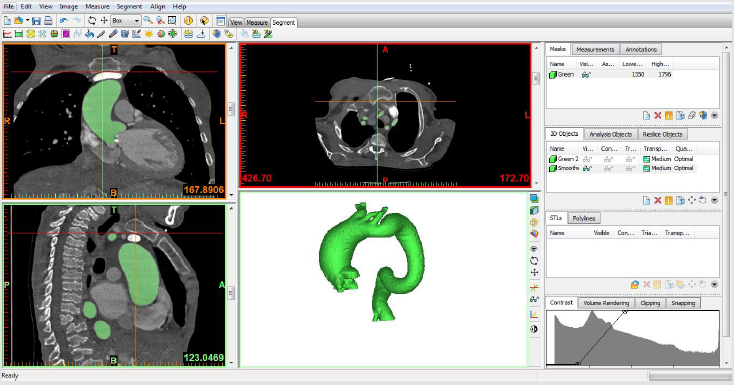
Figure 1: Images showing the segmentation of preoperative CT using mimics.
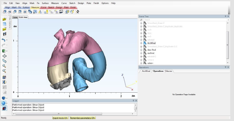
Figure 2: Images showing the generated 3d model using 3d matic.
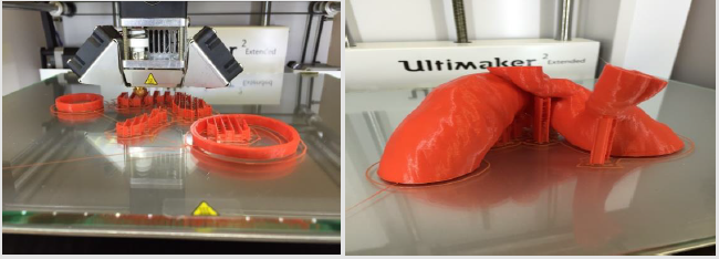
Figure 3: Images showing the printing process of our 3d FDM model.
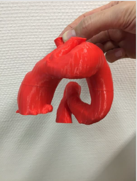
Figure 4: Postoeprative 3d Model
Conclusion
Recently it’s has been shown to be of a great value in preoperative guiding of many surgical interventions. There’s an expanding interests to use patient specific 3D models to guide surgical interventions [2], as well as to generate a bio scaffold to regenerate myocardial tissue [3], critical size bone defects [4] or …. Etc. In cardiovascular surgery it’s yet of limited use in preoperative planning [5], with a great influence in deciding the proper possible surgical intervention. We believe that it offers the patient as well a great chance to understand his/her medical situation and to be integrated in the decision making process. In our case a lot of surgical options were discussed including the endovascular intervention which was at the end declined owing to the good anatomical orientation using the 3d models. We generated a FDM printed 3d model which is to our knowledge would be the first case to use such a technique. The FDM models are to be considered of low cost. Our printed 3d model costs were about (80 euros/ model) which are relevantly low cost compared to other types of 3d printed models [5]. However, it might takes couple of days to build and print the needed model. This technique might not be practical in patients with aortic dissection or other anomalies that needs an emergent intervention. We don’t believe that it should be applied in all patients undergoing routine Interventions. But in case of rare complex anomalies it offers a great help and a good orientation of such rare condition [6].
Disclosure
We had a funding source for this study from the Intramural Research Funding Program of the University Medical Center of the Johannes Gutenberg-University Mainz.
References
- Saric S, Vuletic V, Gvozdanovic V, Mark B (1980) A Case of Kinking of the Aortic Arch. pp. 1147-1149.
- Schmauss D, Haeberle S, Hagl C, Sodian R (2015) Three-dimensional printing in cardiac surgery and interventional cardiology: a single-centre experience. Eur J Cardiothorac Surg 47(6): 1044-1052.
- Leotta DF, Starnes BW (2015) Custom fenestration templates for endovascular repair of juxtarenal aortic aneurysms. J Vasc Surg 61(6): 1637-1641.
- Koleilat I, Jaeggli M, Ewing JA, Androes M, Simionescu DT, et al. (2015) Interobserver variability in physician-modified endograft planning by comparison with a three-dimensional printed aortic model. J Vasc Surg 64(6): 1789-1796.
- Bangeas P, Voulalas G, Ktenidis K (2016) Rapid prototyping in aortic surgery. Interact Cardiovasc Thorac Surg 22(4): 513-514.
- Schmauss D, Juchem G, Weber S, Gerber N, Hagl C, et al. (2014) Three-Dimensional Printing for Perioperative Planning of Complex Aortic Arch Surgery. Case Reports 97(6): 2160-2163.
