 Case Report
Abstract
Breast melanoma metastases are rare and constitute about 1.3-2.7% of cases. In this report we describe a very rare case of metastasis of melanoma of the breast. A 58-yearold woman underwent left mastectomy and axillary dissection for a melanoma metastasis of the breast.
Keywords: Melanoma metastasis; Breast Cancer; Modified Radical Mastectomy; Axillary dissection
Introduction
The incidence of malignant melanoma (MM) is increasing especially in the western population; in Europe, the incidence is less than 10-20 new cases of melanoma per 100,000 inhabitants. MM can spread to other skin sites, lymph nodes or distant organs such as the breast. MM has a great propensity for dissemination and even after primary tumor excision, about 30% of the patients developed metastasis in distant organs [1]. Distant metastases have a poor prognosis with an average survival in untreated patients of only 6-9 months. The excision of one or more metastases is associated with a favorable prognosis for stage IV patients. The presence of metastatic cancer in the breast from other organs is rare but have been described in patients with MM aside from lung carcinoma and carcinoid.
Case Report
A 58-year-old woman visited our Breast Unit outpatient clinic of S.Orsola-Malpighi Policlinic in Bologna, Italy. Two year before she underwent a surgical excision of epithelioid malignant melanoma of the right flank, Clark’s level III, with a negative sentinel lymph node. During follow up multiple pulmonary localizations and a left breast and axillary uptake were found at positron emission tomography (PET). Patient received treatment with Nivolumab with poor effect and dimensional increase of the breast lesion. After multidisciplinary discussion, an attempt with regional radiotherapy was done with progression of the disease. During clinical examination we found a 7 cm ulcerated mass in the upper outer quadrant on the left breast (Figure 1) and pathological palpable lymph nodes in the homolateral axilla. Breast ultrasound showed a suspicious 7cm hypoechoic lesion, with irregular shape of pathological significance in the upper outer quadrants of the left breast. The core needle biopsy (tru-cut) showed melanoma metastasis. The patient underwent a left modified mastectomy (Figure 2) with complete axillary lymph node dissection. Histological report confirmed a melanoma metastasis, S-100, HMB45, Melan-A positive with negative margin status. One out of 28 dissected lymph nodes were positive for macro metastases. One month later patients underwent emergency laparoscopic right hemicolectomy for obstruction. The histological evaluation confirmed ileo-colic metastasis of melanoma. Patient remained relatively stable until she died 8 months after.
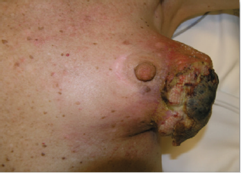
Figure 1: 7 cm ulcerated mass lesion of outer quadrant of the left breast.
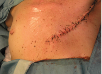
Figure 2: Modified left mastectomy with axillary dissection.
Discussion
Breast melanoma can present clinically as primary melanoma of the breast skin [2], primitive lump of the breast gland [3] or breast metastasis [4]. The most common sites of primary melanoma associated with breast metastases are trunk and upper limb [5]. Because of the good vascularization and the high representativeness of glandular tissue metastasis in the breast occur mainly in the outer quadrants [6]. According to Radvel et al. [6] the average range of onset of the metastases is 52,2 months. In our case the patient presented a breast metastasis 24 months after primary malignancy. A metastatic lesion of the breast is clinically and radiologically similar to a primary breast cancer. Positron emission tomography is not mandatory but may exclude contraindications to surgery [7]. Definitive diagnosis is exclusively histological and immunohistochemistry with particular attention to detect melanoma specific proteins (S-100, HMB45, melan-A). The molecular analysis is mandatory because metastatic melanoma can be treated with checkpoint inhibitors of T cells and BRAF inhibitors. The overall survival rate for patients with stage IV melanoma ranges from 4,7 to 11 months according to the literature [8]. Although with palliative intent, surgical treatment of metastatic breast lumps is the same as for primary breast cancer: quadrantectomy or mastectomy with sentinel lymph node biopsy and additional axillary dissection.
Conclusion
In patients with a personal history of malignant melanoma, metastasis to the breast must be considered when presenting a breast lump.
References
- Essner R, Lee JH, Wanek LA, Itakura H, Morton DL (2004) Contemporary surgical treatment of advanced-stage melanoma. Arch Surg 139(9): 961-966.
- Bono A, Baldi M, Maurichi A, Tomatis S (2003) Distribution of melanoma on breast surface suggests its etiology. Int J Cancer 105: 434.
- Pressman PI (1973) Malignant melanoma and the breast. Cancer 31: 784-788.
- Plesnicar A, Kovac V (2000) Breast metastases from cutaneous melanoma: a report of three cases. Tumori 86: 170-173.
- Al Samaraee A, Khout H, Barakat T, Fasih T (2012) Breast metastasis from a melanoma. Ochsner J 12(2): 149-151.
- Vergier B, Trojani M, De Mascarel I, Coindre JM, Le Treut A (1991) Metastases to the breast: differential diagnosis from primary breast carcinoma. J Surg Oncol 48(2): 112-116.
- Atallah E, Flaherty L (2005) Treatment of metastatic malignant melanoma. Curr Treat Options Oncol 6(3): 185-193.
- Ravdel L, Robinson WA, Lewis K, Gonzales R (2006) Metastatic melanoma in the breast: a report of 27 cases. J Surg Oncol 94: 101-104.
