 Case Report
Abstract
Background: Incidental morphologic findings in lymph node tissue may represent a diagnostic challenge in surgical pathology. We have characterized, for the first time to our knowledge, dense globular structures that most likely represent degenerated lymphoid cells found in a Mascagni-Lund’s lymph node, and resemble Blue Blobs (Bbs) described in vaginal Papanicolaou-stained smears, or Hematoxylin Bodies (Hbs) commonly seen in patients diagnosed with Systemic Lupus Erythematosus (SLE), respectively. These Blue Blob-Like Bodies (Bbbs) are not associated with SLE, and immunohistochemistry may be necessary to rule out malignancy or viral infection.
Objective: To characterize Blue Blob-Like Bodies (Bbbs) in a Mascagni-Lund’s lymph node from a cholecystectomy specimen performed for acute cholecystitis.
Materials and Method: A paraffin-embedded cholecystectomy specimen that included a Mascagni-Lund’s lymph node was evaluated by routine microscopy in a busy surgical pathology service. Special stains, immunohistochemistry, and electron microscopy were performed to characterize the unusual structures observed.
Results: Many scattered, round-to-oval, dense globular basophilic novel structures with smooth borders were identified and named Blue Blob-Like Bodies (Bbbs).
Conclusion: Bbbs mimic malignant (primary or metastatic) or virally infected cells in lymph node tissue. However, they most likely represent degenerated lymphoid cells resulting from reactive lymphadenopathy secondary to acute cholecystitis.Pathologists need to recognize Bbbs and properly distinguish them from other pathological structures, in order to avoid misdiagnosis.
Keywords: Blue Blobs; Blue Blob-Like Bodies; Electron Microscopy; Hematoxylin Bodies; Immunohistochemistry; Lymph Node Tissue; Malignancy; Mascagni- Lund’s Lymph Node
Abbreviations: BBs: Blue Blobs; HBs: Hematoxylin Bodies; SLE: Systemic Lupus Erythematosus; BBBs: Blue Blob- Like Bodies; PAS: Periodic Acid-Schiff; EMA: Epithelial Membrane Antigen; CEA: Carcinoembryonic Antigen; DNA: Deoxyribonucleic Acid; MGP: Methyl Green-Pyronin;
Introduction
Incidental morphologic findings in lymph node tissue may represent a diagnostic challenge in either benign or malignant surgical pathology. While malignancy could be primary or metastatic, the etiology of benign pathology ranges widely and includes infectious, inflammatory, and autoimmune disease. For instance, virally infected cells may be found serendipitously, especially in immunosuppressed patients. Similarly, various pathologic bodies have been described in different tissues, including lymph nodes. In 1932, Gross [1] described for the first time, amorphous extracellular deposits of basophilic material that could be stained with hematoxylin in patients with endocarditis due to Systemic Lupus Erythematosus (SLE). These structures, known as Hematoxylin Bodies (Hbs), are considered highly specific for SLE. In 1959, Pollack [2] recognized Hbs in lupus lymphadenitis. Soon after, Candreviotis[3] described Hbs-like material in nodal tissue from a patient with Classical Hodgkin Lymphoma, a finding which has not been reproduced yet. In addition, Handra-Luca et al. [4] noted the presence of lipogranulomata in a sentinel gall bladder lymph node (Mascagni’s or Lund’s node), which may morphologically suggest metastatic signet ring adenocarcinoma. Therefore, characterization of unique structures that could be confused with atypical cells (malignant or virally infected) or known pathologic bodies remains significant. For example, the so-called blue blobs (Bbs) described by Milligan et al. [5] may be a source of misdiagnosis in postmenopausal vaginal smears, since they simulate malignancy. Likewise, a few structures such as crystals or Liesegang rings have been confused with parasitic organisms and may be associated with malignancy [6,7]. Herein, we describe previously unrecognized atypical material in a Mascagni-Lund’s lymph node from a specimen obtained after cholecystectomy for acute cholecystitis.
Materials and Methods
A cholecystectomy specimen that included a Mascagni-Lund’s lymph node was obtained from a 59-year-old male smoker. The patient, who had previous medical history of hepatitis C (status post-treatment completion) and hypertension, was admitted to the hospital with clinical and laboratory diagnosis of acute cholecystitis. The cholecystectomy was performed 72 hours following clinical symptoms and antibiotic treatment. The specimen was fixed in formalin, embedded in paraffin, and prepared for evaluation by routine microscopy. Histopathology revealed chronic cholecystitis and cholelithiasis, and a 1cm Mascagni-Lund’s lymph node with unusual structures resembling large bare nuclei. Special stains, immunohistochemistry, and electron microscopy were performed to characterize these structures.
Results
Hematoxylin and Eosin (H&E) stained sections from paraffinembedded tissue revealed a small lymph node with preserved architecture and follicle lysis, containing naked germinal centers. Scattered round-to-oval dense globular basophilic structures with smooth borders, resembling cells with large atypical nuclei and high Nuclear/Cytoplasmic (N/C) ratio, were identified in the interfollicular areas and sinuses (Figure 1). These non-polarizable structures were suspicious for malignancy or viral infection. Morphologically they best resemble blue blobs (Bbs), as delineated by Abdulla et al. [8] and are henceforth referred to as blue bloblike bodies (Bbbs). Bbbs varied in size from 5-30 μ, and showed homogenous dark blue color and somewhat coarse texture (Figure 2). Special stains revealed Bbbs to be positive for Feulgen (Figure 3), and negative for calcium, mucicarmine, Periodic Acid- Schiff (PAS) and Romanowsky-Giemsa. Immunohistochemistry revealed the Bbbs to be partially positive for CD45 (Figure 4), and negative for Pankeratin AE1/AE3, S100, PAX-5, CD20, CD3, CD56, CD31, CK8/18, Epithelial Membrane Antigen (EMA), monoclonal Carcinoembryonic Antigen (CEA), herpes antigens (HSV1 and HSV2), cytomegalovirus and Ki-67. Electron microscopy showed acellular, globular material varying in size from 5-30 μ, which is compatible with degenerating cells.
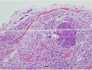
Figure 1: Low power H&E stain (200X) reveals lymph node with few naked germinal centers and scattered Bbbs mimicking large atypical nuclei.
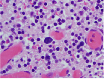
Figure 2: High power H&E stain (1000X) shows basophilic homogenous dark Bbbs with somewhat coarse texture.
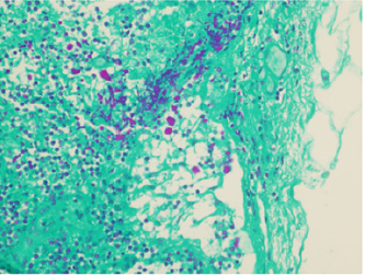
Figure 3: Feulgen stain demonstrates positivity on Bbbs (400X).
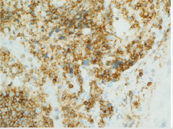
Figure 4: CD45 immunostain highlights partial positivity on Bbbs (400X).
Discussion
To our knowledge, this is the first description of Bbbs found in lymph node tissue. Bbbs appear to have no clinical significance. Nonetheless, they are pathologically intriguing since they can be potentially misinterpreted and misdiagnosed as malignancy, viral infection, or other previously described histopathologic structures, such as Bbs or Hbs. Bbbs very closely resemble Bbs, as described first on Papanicolaou-stained vaginal smears in 1959 by Milligan et al. [5] and further characterized in 1999 by Abdulla et al. [8] Although the two bodies bear close similitude, characteristic location, morphology and staining profile (special and immunohistochemical) allows a clear distinction. For instance, Bbbs reside in lymph node tissue and show higher size variability ranging from 5-30 μ, as opposed to the 8-15 μ described for Bbs, which are found in vaginal smears. Interestingly, both bodies are composed mostly of Deoxyribonucleic Acid (DNA), as demonstrated by positivity for Feulgen stain. However, Bbs are also positive for PAS, Methyl Green-Pyronin (MGP), cytokeratins (CAM 5.2, AE1/ AE3), EMA and CEA, while Bbbs are only positive for CD45, and negative for the rest. These findings suggest that both Bbs and Bbbs are DNA-rich structures likely representing degenerated cells, which have distinct epithelial and hematopoietic origins, respectively.Also, Bbbs could be mistaken for Hematoxylin Bodies (Hbs) since both contain degenerated nuclear material, as proven by positive Feulgen staining. However, significant differences between these two structures exist. For example, Hbs are typically found in patients with SLE, [3,9] although a single report described them in association with Hodgkin Disease [3]. Nevertheless, the illustrations of Hbs in this historic publication have been considered unconvincing, [9] and therefore, the strong association of Hbs and SLE remains valid. Interestingly, our patient does not have current or past medical history of SLE, other autoimmune disease, or malignancy. Moreover, Hbs are commonly present in necrotic tissue including necrotizing lymphadenitis, [10] but necrosis was absent in the gallbladder neck lymph node analyzed here. Furthermore, on H&E stains, Hbs display larger size, more angulated shape, and rougher edges than Bbbs, which are smaller, smoother, and rounder. Finally, Hbs are amphophilic (magenta color) on Romanowsky-type stains, while Bbbs are basophilic. Consequently, although Hbs and Bbbs may both represent cellular degeneration, they are dissimilar. In summary, Bbbs are novel DNA-rich globular structures, which seem to represent degenerated lymphoid cells resulting from reactive lymphadenopathy secondary to acute cholecystitis. Bbbs can be differentiated from Hbs based on their histopathological features and disassociation with SLE. Likewise, Bbbs must be distinguished from other atypical structures such as malignant or viral infected cells, to prevent misdiagnosis during the practice of surgical pathology.
Acknowledgment
Sincere gratitude to Mrs. LyvouchFilkoski for excellent technical assistance.
Disclosure
Funding Sources: No conflict of interest to disclose.
References
- Gross L (1932) The heart in atypical verrucous endocarditis (Libman-Sacks). In: Contributions to the medical sciences in honor of Dr. Emanuel Libman by his pupils, friends, and colleagues. The International Press, New York, NY, USA, 2: 527-550.
- Polack AD (1959) Some observations on the pathology of systemic lupus erythematosus. J Mt Sinai Hosp N Y 26: 224-240.
- Candreviotis N (1962) Hematoxylin bodies in Hodgkin’s disease. J Clin Pathol 15: 542-543.
- Handra-Luca A, Ben Romdhane MH, Straub BK (2016) Lipid histiocytosis of the gallbladder neck lymph node. Case Rep Pathol 2016(3): 1-4.
- Milligan M, Carrow LA, Eggers V (1959) A source of false positives in cytologic interpretation. Am J Obstet Gynecol 78: 599-603.
- Tao Q, Zhang W, Chen Z, Gao L, Yan J, et al. (2019) Generalized crystal-storing histiocytosis with diffuse large B-cell lymphoma and monoclonal gammopathy in a Chinese elderly woman: a case report. BMC Cancer 19: 514.
- Sancheti S, Jain S (2017) Liesegang rings: Extremely rare structures in malignant lesions. Int. J Surg Path 26: 39-40.
- Abdulla M, Hombal S, Kanbour A, Becich M, Stankovich D, et al. (2000) Characterizing “Blue Blobs”: Immunohistochemical staining and ultrastructural study. Acta Cytol 44: 547-550.
- Mc Mullan DT, Powers JM, Nussbaum AI (1988) Hematoxylin-stained bodies and tissue LE cells in a skeletal muscle biopsy. Am J Clin Pathol 90: 731-733.
- Indu RN, Suma B, Pooja P, Menila D (2020) Clinicopathologic spectrum of necrotizing lymphadenitis. Indian J Pathol Microbiol 63: 60-63.
