 Research Article
Abstract
In order to study the role of the amygdala-hypothalamic relationship in the organization of the hippocampal theta rhythm, we studied the amplitude-frequency analysis of the EEG of the dorsal (CA1 and CA3 fields), ventral hippocampus, dentate fascia and medial nucleus of the septum under conditions of electrical stimulation and electrolytic destruction of DAP and VAP. The data obtained indicate a differentiated effect of DAP and VAP on the mechanism of the formation of the hippocampal theta rhythm, as well as the need for functional integrity of DAP, which is one of the key links in the mechanisms of regulation of neuronal excitability in the septo-hippocampal system.
Keywords: Dorsal and Ventral Amigdalofugal Ways; Spectrogram of the Hippocampus; Amplitude-Frequency Analysis; Electrostimulation; Destruction
Abbreviations: DAP: Dorsal Amigdalo-Fugal Pathway; VAP: Ventral Amygdalo-Fugal Pathway; ABE: Amygdale; MFB: Medial Forebrain Bundle
Introduction
Numerous studies have shown the presence of bilateral connections between the amygdala and the hypothalamus. Bilateral monosynaptic connections of the amygdala with hypothalamic formations and other structures of the brain stem are carried out by two systems of amygdala fibers: dorsal amigdalo-fugal pathway (DAP) and ventral amygdalo-fugal pathway (VAP) [1]. DAP or ST (stria terminalis) - a compact bundle of fibers - contains amygdalofugal and amygdalopetal pathways. The main part of the ST afferent fibers passes through the central (AC) and basolateral (AB) nuclei of the amygdala. Several scattered beams go to medial nucleus of the amygdale (AME) and through it to cortical nucleus of the amygdale (ACO) [1]. At the level of the anterior hypothalamus, the fibers enter the medial forebrain bundle (MFB), and at the level of the preoptic zone, a massive distribution of the postcommissural component of the ST is observed. Here the ST fibers overlap with the VAP fibers. A part of the projection zone of the dorsal DAP component is the amygdalo-hippocampal transition zone [2], which confirms the presence of a connection between DAP and MFB [3]. This group of fibers, belonging to the DAP, provides interaction with the fibers of the ventral tract from the amygdala, which form the VAP. The two main systems of amygdala fibers described by morphologists - DAP and VAP - provide bilateral monosynaptic connections of the amygdala primarily with hypothalamic formations and other structures of the brain stem. Diffuse fibers of VAP are less studied. These ventral fibers terminate in the core of the ST bed, in the septum, the preoptic region, and in the anterior hypothalamus. Their projection area therefore partially overlaps with the ST area [3]. To investigate the role DAP and VAP in the organization of the theta rhythm of the hippocampus, we studied the amplitude-frequency analysis of the EEG of the dorsal (fields CA1 and CA3), ventral hippocampus, dentate fascia and medial nucleus of the septum under conditions of electrical stimulation and electrolytic destruction of DAP and VAP.
Methods
Experiments were carried out on 20 mature rabbits weighing 2.5-3.0kg. The EHipG was recorded from the dorsal hippocampus (the CA1field: P 3.0, L 2.0, H 18.0, and the CA3 field: P 0 2.0, L6.0, H17.0) and from the medial nucleus of the septum (A-3.0; L0.5; H10.5) on the encephalograph Medikor EEG-16E with the use of needle electrodes insulated except the tip. Spectral EHipG analysis was performed using standard electroencephalographic approaches. Electrostimulation and electrical destruction of the dorsal - DAP (precompassural region: A-1; L3.2: H11.5) and ventral- VAP (P5; L6H15.8) amygdalofugal pathways were made by bipolar electrodes with currents of up to 1.0 mA for 15–25 sec. A histogram method was used for amplitude-frequency analysis of the EEG, as described by Fujimori [4].
Results and Discussion
After registration of the background activity, electrical stimulation of the dorsal and ventral amygdalofugal pathways was performed. Unilateral stimulation of both the DAP and VAP at low parameters of the irritating current (50-80μA, frequency 10imp/s, with a stimulus duration of 0.5msec) leads to significant changes in the electrical activity of the studied brain structures. Frequency analysis of the EEG of the ipsi- and contralateral hippocampus clearly showed that if in the background the activity was characterized by the predominance of theta waves in the range of 4-6 oscilation/ s, then at the moment of electrical stimulation of the DAP an increase in the frequency of the theta rhythm was observed up to 6-7.5 oscilation / sec from. At the same time, a decrease in the representation of fast-frequency beta oscillations was noted. Amplitude analysis revealed an increase in the EEG amplitude of the ipsi- and contralateral sides of the dorsal (fields CA1 and CA3), ventral hippocampus, dentate fascia and medal nucleus of the septum (Figures 1AI & 1AII). Further increase in stimulation parameters (100-200μA, frequency 20 pulses / s with a stimulus duration of 0.5msec) for 10-15 seconds from the first minutes of exposure in the electrical activity of all registered structures, simultaneously synchronized activity at 2-5 seconds of stimulation turns into epileptiform.
Over time, epileptiform activity completely disappears, and a flattened, low-amplitude activity is recorded in the hippocampus, well pronounced on the ipsilateral side of destruction. It is quite significant that the effect of suppressing theta activity in the hippocampus after DAP damage was irreversible: its recovery was not observed even 6 months after the coagulation. Unilateral electrolytic destruction of DAP led to complete and irreversible blockade of the hippocampal theta rhythm. It is quite significant that the effect of suppressing theta activity in the hippocampus after DAP damage was irreversible: its recovery was not observed even 6 months after the coagulation. Analysis of the spectrogram of the hippocampus after the destruction of the DAP revealed the complete disappearance of theta rhythm and the dominance of slow alpha waves and fast beta oscillations. Amplitude analysis showed a sharp decrease in the amplitude of EEG activity (Figures 1BI & 1BII). In contrast to the damage to the DAP in unilateral electrolytic damage to the VAP, there was no complete disappearance of theta waves. In the hippocampus, irregular, deformed, polymorphic activity was recorded, combining both slow and individual theta waves, however, the main part of the EEG is made up of highfrequency beta oscillations.
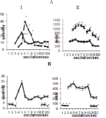
Figure 1:
A. Amplitude-frequency analysis of the electrical activity of the ipsilateral hippocampus before and after electrical stimulation and
B. Electrolytic destruction of the dorsal amygdalofugal pathway. I - frequency, II - amplitude analysis. I - frequency distribution polygon. The abscissa is the average value of the classes of intervals in frequencies, the ordinate is the frequency of repetition; II - amplitude-frequency characteristic.Solid line - before, dashed - after exposure.
In the case of damage to the VAP, in contrast to the destruction of DAP, there is a complete restoration of the EEG activity of the studied brain structures to the background level on the 20-25th day. The performed amplitude-frequency analysis showed the presence of sigma waves that were absent in the background, a decrease in the representation of theta waves, and a sharp increase in fastfrequency oscillations. Amplitude analysis revealed a decrease in the amplitude of the activity of the studied brain structures (Figures 2AI & 2AII; 2BI & 2BII). The possibility of modulating the electrical activity of the hippocampus and especially its theta rhythm by changing the functional state of the extrahippocampal structures of the brain, including the hypothalamus and amygdala, is well known [5-8]. The fundamental difference between our data is the fact that under conditions of coagulation of DAP, there is not modulation, but a deep and long-term depression of the total electrical activity of this structure. Considering the regulatory effect of the amygdala on the functional state of hypothalamic neurons [9], as well cells of the hypothalamus [3], it can be assumed that with weak stimulation of DAP and VAP, hypothalamic- pituitary adrenal system is activated, which results in the release of adrenergic corticotropic and steroid hormones in doses leading to the registration of synchronized highamplitude activity in the hippocampus.
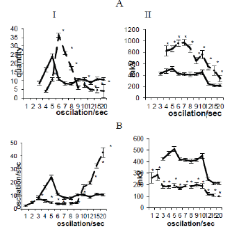
Figure 2:
A. Amplitude-frequency analysis of the electrical activity of the hippocampus before and after electrical stimulation and
B. Electrolytic destruction of the ventral amygdalofugal pathway. I - frequency, II - amplitude analysis. Solid line - before, dashed - after exposure.
Increasing stimulation parameters DAP and VAP promotes an increase in the concentration of hormones in the blood, which leads to an increase in the excitability of hippocampal neurons and registration of epileptiform activity. The destruction of DAP, in contrast to VAP, leads to irreversible changes in the EEG of the hippocampus. Since the hippocampus and septum are the main targets for corticosteroids, which regulate the excitability of nerve cells, then after the destruction of the ST, the hypothalamus is obviously out of control of the amygdala, the activity of the HPA is disrupted and, as a result, the amount of adrenergic corticotropic and steroid hormones in the blood increases, which during 2-3 hours cause hyperactivity (epi-discharges) of hippocampal neurons, followed by the death of pyramidal cells of the hippocampus, which is reflected in the blockade of activity. All of the above indicates the differentiated effect of DAP and VAP on the formation mechanism of the hippocampal theta rhythm, as well as the need for the functional integrity of DAP, which is one of the key links in the mechanisms of regulation of neuronal excitability in the septo-hippocampal system.
References
- Chepurnov SA, Chepurnova NE (1981) Almond-shaped brain complex. M Publishing house, Mosk University, Morocco, pp. 256.
- Post St, Mai JK (1980) Contribution to the amygola loid projection field in the rat. A quatitative autoradiographic study. J Hirnforsch 21(2): 199-225.
- Sapronov NS (1998) Pharmacology of the hypophyseal-adrenal system. Publishing house "Special. literature ", St. Petersburg. p. 336.
- Fujimori B, Yokota T, Ishibashi Y, Takei T (1958) Analysis of the electroencephalogram of cjildren by histoqram method. EEG and Clin Neurophysiol 10(2): 241-252.
- Kitchigina VF (2006) Regulation mechanisms and functional significance of theta oscillations in the septohippocampal system of the brain. Abstract of dissertation for the degree of Doctor of Biological Sciences, Moscow, Pushchino.
- Kitchigina V, Popova I, Sinelnikova V, Malkov A, Astasheva E, et al. (2013) Disturbances of septohippocampal theta oscillations in the epileptic brain: Reasons and consequences. Experimental Neurology 247: 314-327.
- Steriade M (1996) Arousal: revisiting the reticular activating system. Sciense 272(5259): 225-226.
- Vertes RP (1992) PHA-L analysis of projections from in the supramammillary nucleus in the rat. J Comp Neurology 326(4): 595-620.
- Olmos JS De, Ingram VR (1972) The projection field of the stria terminals in the rat brain. An experimantal study. J Compar neurol 146(3): 303-315.
