 Case Report
Synopsis
A nine years old cross-breed Holstein Friesian dairy cow weighing 280kg with medium body condition was presented to Mekelle University Veterinary Teaching Hospital Mekelle, Ethiopia. The cow was presented with the signs of frequent abdominal distention, abdominal pain, grunting, grinding of teeth, reduced feed intake, progressive weight loss, and falling behind as the owner complains. The detailed clinical examination revealed that dull, depressed, reduced appetite and weakness, and the case was confirmed as traumatic reticuloperitonitis. The case was handled surgically by exploratory rumenotomy after preparing the surgical site was prepared aseptically and stabilizing with fluids and electrolytes preoperatively. Para-vertebral nerve block by using 2% lignocaine hydrochloride was done. A long skin incision starting about eight to ten centimeters below the transverse process of the lumbar vertebrae was made vertically. The abdominal muscles and peritoneum were also incised following the skin incision. The rumen was fixed with the stay suture and metallic foreign bodies were removed using magnetic material through exploration. After removing the content of the rumen, it was closed by double lambert suture. Then closing the peritoneum and muscle with catgut by simple continuous pattern. The skin was sutured with nylon using a cross mattress suture. The cow was completely recovered after 20 days of followup observation. In the current case report, the successful management of traumatic reticuloperitonitis due to metallic foreign bodies in a cow is discussed.
Keywords: Dairy Cow; Metallic Foreign Bodies; Exploratory Rumenotomy
Introduction
Ingestion of foreign bodies by ruminants is extremely common
especially in developing countries, like Ethiopia, where the standard
of animal management is unsatisfactory, and low nutritional statuses
of ruminants have forced them to scavenge for food [1]. Rapid
urbanization, industrialization, and acute mineral deficiencies
are that predispose them for foreign body ingestion. Accordingly,
they cause great production loss and decrease reproductive
performance [2]. Ruminants frequently ingest irregular objects with
potential risk of rumeno-reticular damage. Non-metallic foreign
bodies, plastic foreign bodies, ropes, and pieces of old clothes are
commonly ingested by cattle and accumulated in the reticulorumen
causing a variety of ailments [3,4].Traumatic Reticuloperitonitis
(TRP), also called hardware disease, is a relatively common disease
in adult cattle that impairs reticuloruminal motility. It is caused
by the ingestion and perforation of the reticular wall resulting in
acute perireticular inflammation followed by peritonitis, pleuritis,
and/or pericarditis, in addition to sepsis, restrictive adhesions, and
abscess formation., Perireticular adhesions, and abscesses [5,6].
The condition is usually caused by long, thin, sharp foreign bodies
(wire, needles, nails) that penetrate the reticulum, peritoneum,
diaphragm, and pericardial sac, eventually leading to traumatic
pericarditis. This leads to inflammation of the pericardium, with
the accumulation of serous or fibrinous inflammatory products
[7]. The influencing factors of TRP include remodeling of livestock
housing, careless handling of baling wires, pins, feed sack bags,
and wires. The incidence is more in females than males shortly
after calving [8,9]. Most cattle owners with hardware disease often
present with complaints of rumeno-reticular dysmotility (bloat),
abdominal discomfort (colic), anorexia, lethargy, and weight
loss (falling behind). Recurrent rumen tympany, pale mucous membrane, rumen impaction, atony, reduction of rumen motility,
scanty faces, partial anorexia, labored breathing, grunting, weight
loss, and colic are common clinical signs of TRP [7,10].
Evidence of pain localized to the cranioventral abdomen, ruminal atony during auscultation, and pleural/peritoneal/ pericardial effusion during percussion should be performed during physical examination. Additionally, inflammatory leukogram, X-ray (detection of the presence of foreign body), and ultrasonography (characterizing pericardial effusion as well as determine the extent of the lesions and assess the prognosis) are best and preferred methods of diagnosis for before exploratory laparotomy or rumenotomy. Even though prevention should be the primary emphasis, the surgical management of hardware disease should aim at controlling infection and removing foreign bodies [1,4]. The present case report describes the history, signalment, physical, and surgical findings and result in adult dairy cattle following exploratory rumenotomy.
Case Report
Case History and Clinical Examination
A multiparous nine-years-old cross-breed Holstein Friesian dairy cow weighing 280 kg with a medium body condition was presented to Veterinary Hospital, College of Veterinary Science (CVS), Mekelle University (MU), Ethiopia. The owner also told as the cow was suffering from grunting, grinding of tooth, abdominal pain, reduced feed intake, progressive weight loss, frequent abdominal distention, and falling behind. Besides, the cow graze extensively on the nearby premises and decreased milk production over the last three months. Upon arrival and physical examination, the cow was lethargic and highly depressed with a rough hair coat and dehydrated. Further close examination of vital organ parameters such as heart rate, respiratory rate, pulse rate, and mucous membrane revealed slightly elevated. Besides, upon poll and pinch grip test, the cow showed signs of pain, discomfort. Moreover, the cow was having difficulty in regurgitation, ruminal atony up auscultation, and intermittent straining at frequent intervals. Accordingly, based on the history and clinical observation, the case was diagnosed as traumatic reticuloperitonitis and the team decided to manage surgically using exploratory rumenotomy.
Animal Handling, Anesthetic Protocol, and Preoperative Patient Preparation
Preoperatively, the cow was kept on antibiotics for three consecutive days and fluid therapy (5% Dextrose solution plus 0.9% NaCl, 1000ml stat, I.V., Addis pharmaceutical, Adigrat Ethiopia) to correct the dehydration status. The cow then was properly restrained with the combination of physical (using rope) and chemical methods (Detomidine hydrochloride, Dechra Veterinary Products Ltd., the United Kingdom at a dose of 20μg/kg Intravenously). Then, the skin surface on the left paralumbar fossa was prepared aseptically by washing with water, soap, and salvon® (Cetrimide 3% and Chlorhexidine gluconate 0.5% solution). Then the hair was first clipped with sharp scissor and shaved with a razor blade and cleaned thoroughly with a standard solution of salvon®. Finally, the area was scrubbed three times with a povidone-iodine 1% solution to decrease the microbial load in the area and left dried till ready for Rumenotomy (Figure 1A).
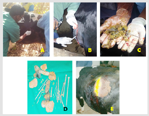
Figure 1: Surgical procedure during Rumenotomy in Crossbreed Holstein Friesian dairy cow
A) Physical restraining and aseptic preparation of the cow on standing position using rope.
B) A sharp vertical incision on the skin and abdominal muscle of the left flank area.
C & D) Presentation of metallic foreign bodies taken out from the Rumen.
E) Post-surgical appearance of the crossbreed Holstein Friesian dairy cow.
Additionally, regional or field block was performed by infiltrating the 5 ml lidocaine per each paravertebral space (Lidocaine hydrochloride 2%, Vedco Inc. Saint Joseph Missouri) to block T13, L1 and L2 nerves using an 18-gauge syringe having a 10-cm needle, to desensitize the flank area, abdominal muscles and alleviate pain during the surgical procedure. The needle was inserted halfway between the intervertebral transverse process and the needle is slightly angled to reach and deposit the lidocaine in the subarachnoid space. Finally, two linear infiltrations were made in the pattern of inverted ‘T’ using local anesthetic lidocaine (60ml) to desensitize and put in sufficient analgesia enclosing the site of incision and waited for 10-minutes.
Surgical Correction
Abdominal surgery such as rumenotomy is a cleancontaminated
operation. For the rumenotomy, an operation
room is necessary. However, as there was no operation room we
operated outside. A sharp vertical skin incision with a distance of
approximately (~40 cm long) was made on the left flank region
below the lumbar transverse process (Figure 1B). After blunt
dissection of the skin from the subcutaneous tissue, the incision
was continued chronologically through the external and internal
abdominal oblique, transverse abdominal muscle, and peritoneum.
Then all muscular layers together with skin were grasped with
handheld retractor to get sufficient surgical field and exposure to
the rumen. Stay suturing of the rumen was performed to fix. After
the incision of the rumen, exploration of the rumen and reticulum
was conducted and different metallic, as well as plastic foreign
bodies, were removed (Figure 1C &1D).
Moreover, bleeding during the procedure was managed
by applying sterile gauze, using different straight and curved
hemostatic forceps and topical infiltration of epinephrine on
bleeding site depending on the site and condition. Then, the rumen
and surrounding area were rinsed copiously with sterile isotonic
saline solution and is closed by double lambert suture using sterile
absorbable polyglycolic acid of size 1-0 (Shandong Sinorgmed
Int’l Co., Ltd, China) and replaced in the abdomen to its normal
position. The peritoneum was closed by simple continuous suture
and muscle with a continuous lockstitch pattern using 2-0 size
sterile absorbable polyglycolic acid. Then subcutaneous suture
and skin suture for closing the skin. After that, the skin together
with subcutaneous facia was closed with horizontal interrupted
mattress using silk (2-0 size). Lastly, the area was properly cleaned
and dressed in a 2% povidone-iodine solution and admitted home.
Post-Operative Follow Up and Result
Postoperatively, small skin bleeding was noticed but it is normal and helps to heal the wound unless continued for a long period. Antiseptic wash of the incised area was done at second- and thirddays post-operative up to fourteen days until it completely healed. The suture was removed after 14days. Besides, the cow was kept on fluid therapy (5% Dextrose solution, 1000ml stat, I.V.) antibiotic (Ceftriaxone, 5gm/kg, I.V.), and Meloxicam (0.5mg/kg, IM) for five successive days. Tetracycline wound spray was also applied around the wound (Figure 1E).The owner was also advised to closely monitor the cow and to provide good nutrition to facilitate wound healing. After 20-day post-operation, the wound was healed completely and after two months of follow-up, the cow was under good health status (Figure 1).
Discussion
Surgical affections of the ruminant forestomach due to ingested foreign bodies are the subject of attention almost all over the world and of major economic importance due to severe loss of production and production ability [1,2,4]. These findings agree with the current cases in that there were weight loss and debilitation due to the presence of plastic foreign bodies in the cow’s rumen.In the present case report, emaciation, pale mucus membrane, scanty feces, anorexia, rough hair coat, and distended abdomen were among the common clinical findings in the affected cow. These results were in agreement with the report by [1,4,11] in which animals with indigestible foreign body cannot able to digest feeds normally so that prone to feed deficiency which can be manifested by different clinical signs. Besides, most of the foreign bodies were removed from the rumen while the rest were removed from the reticulum. This finding was in line with the findings of [1,2,12] that may result in enlargement of the rumen.
Rumenotomy is the most preferable method of removing
metallic and plastic foreign bodies from the rumen and reticulum.
It is a clean-contaminated surgery since a hollow viscous is
penetrated. Wound dehiscence, hemorrhage fever, edema, wound
infection (peritonitis), death, intestinal obstruction/adhesion, and
physiological bloat are among the commonly observed postoperative
complications. Post-operative care should be emphasized through
the administration of antibiotics and analgesics to minimize the risk
of post-operative complications [8,13]. This was inconsistent with
the current case report in terms of case handling, treatment, and
post-operative care.Surgical removal of foreign bodies improved
the body condition, feed intake, productivity, and wellbeing of the
cow. This finding agreed with an experimental study conducted by
[14] who concluded that the surgical removal of the foreign body
did improve the health of the animals by increasing the feed intake,
weight gain, and productivity following removal of the foreign body.
In Ethiopia, the extensive management system, shortage
of forage during the dry season, and nutritional deficiency are
considered as a major predisposing factor for acquiring indigestible
rumen foreign bodies in ruminants. This resulted in a serious health
impact on the cattle and a threat to the environment [1,2,4,8].
Similarly, the findings of the current study revealed the frequent
occurrence of rumen foreign bodies in cattle. The indiscriminate
ingestive behavior of cattle [9] as compared to small ruminants could be given as an explanation for the higher prevalence in this
species. Moreover, widespread use and improper disposal of the
non-bio-degradable free polythene or plastic shopping bags for
packaging and other garbage and the poor waste management
system in the country and shortage of animal feed could be reasons
for the occurrence of the problem in ruminants [1,12].
Conclusion
The cattle owner should be advised to give a balanced type of feed to minimize the risk of ingesting foreign materials due to mineral deficiency. Besides, the movement of the animals should be restricted to avoid the predisposition to metallic and plastic foreign bodies. The owner should understand the outcome of the disease. So, recommend that once the case occurred it should have to bring the animals as early as possible before it is emaciated and immunologically compromised.
References
- Kebede S, Bekele T, Fesseha H (2020) Prevalence of Indigestible Rumen and Reticulum Foreign Bodies in Cattle Slaughtered at KombolchaElfora Abattoir, Kombolcha Town, Amhara Regional State, Ethiopia. Int J Rec Biotech 8(1): 25-34.
- Ramaswamy V, Sharma HR (2011) Plastic bags-threat to environment and cattle health: A retrospective study from Gondar city of Ethiopia. IIOAB J 2(1): 6-11.
- Bassa K, Tesfaye W (2017) Study on rumen and reticulium foreign bodies in cattle slauthered at WolaitaSodo municipal Abattoir, Ethoipia. Int J Adv Multidiscip Res 4(1): 11-19.
- Mekuanint S, Alemneh T, Asredie T (2017) Indigestible rumen foreign bodies-causes rumen impaction in cattle, sheep and goats slaughtered at Addis Ababa Abattoir Enterprise, Ethiopia. Journal of Veterinary Science and Medicine 5(1): 5.
- Fubini S, Ducharme N, Erb H, Smith D, Rebhun W (1989) Failure of omasal transport attributable to perireticular abscess formation in cattle: 29 cases (1980-1986). Journal of the American Veterinary Medical Association 194(6): 811-814.
- Rehage J, Kaske M, StockhofeZurwieden N, Yalcin E (1995) Evaluation of the pathogenesis of vagus indigestion in cows with traumatic reticuloperitonitis. Journal of the american veterinary medical association 207(12): 1607-1611.
- Braun U, Lejeune B, Schweizer G, Puorger M, Ehrensperger F (2007) Clinical findings in 28 cattle with traumatic pericarditis. Veterinary Record 161(16): 558-563.
- Anteneh M, Ramswamy V (2015) Hardware disease in bovine. Acad J Anim Dis 4(3): 146-159.
- Reddy Y, Latha P, Reddy S (2014) Review on metallic and non-metallic foreign bodies: A threat to livestock and environment. Int J Food Agric Vet Sci 4: 6-14.
- Chanie M, Tesfaye D (2012) Clinico-pathological findings of metallic and non-metallic foreign bodies in dairy cattle: A review. Acad J Anim Dis 1(3): 13-20.
- Bakhiet AO (2008) Studies on the rumen pathology of Sudanese desert sheep in slaughter house. Sci Res Essays 3(7): 294-298.
- Tiruneh R, Yesuwork H (2010) Occurrence of rumen foreign bodies in sheep and goats slaughtered at the Addis Ababa Municipality Abattoir. Ethiopian Veterinary Journal 14(1): 91-100.
- Fubini S, Ducharme N (2016) Farm Animal Surgery-E-Book. Elsevier Health Sciences.
- Ghurashi M, Seri H, Bakheit A, Ashwag E (2009) Effect of surgical removal of foreign body from goat’s rumen with special reference to the prevalence of foreign body in goats in Southern Darfur. Australian Journal of Basic and Applied Sciences 3(2): 664-668.
