 Research Article
Abstract
We demonstrated that nitrous acid gas (HONO) which is measured as nitrogen dioxide (NO2), induced pulmonary emphysema-like alterations in guinea pigs after continuous exposure for 4 weeks at 3.6 Parts Per Million (ppm), and we suggested that HONO affects asthma symptoms more than NO2 according to a 5.8-ppm-HONO exposure experiment measuring respiratory function in rats. This study aimed to investigate the dose–response effects of HONO exposure on histopathological alterations in the respiratory tract of guinea pigs to find the Lowest Observed Adverse Effect Level (LOAEL) of HONO. We continuously exposed male Hartley guinea pigs (n = 5) to four concentrations of HONO (0.0, 0.1, 0.4, and 1.7 ppm) for 4 weeks (24 h/day). We performed histopathological analyses: observation under a light microscope including measurement of the alveolar Mean Linear Intercept (Lm) and the thickness of the bronchial smooth muscle layer for three guinea pigs in each group, and observation under a Scanning Electron Microscope (SEM) and a Transmission Electron Microscope (TEM) for two guinea pigs in each group. We found pulmonary emphysema-like alterations in the centriacinar regions of alveolar ducts, a significant increase of Lm in the 1.7-ppm-HONO-exposure group, a tendency for hyperplasia and pseudostratification of bronchial epithelial cells, and the extension of the bronchial epithelial cells and smooth muscle cells in alveolar duct regions. These results had a dose-dependent tendency. The present results suggest that the LOAEL of HONO is < 0.1-ppm by histopathological analyses.
Keywords: Nitrous Acid; Nitrogen Dioxide; Pulmonary Emphysema; Alveolar Mean Linear Intercept; Asthma
Introduction
Numerous epidemiological studies have shown the relationship between nitrogen dioxide (NO2) and the alterations of respiratory function or asthma symptoms [USEPA, 2016]. However, conventional NO2 assays measure nitrous acid (HONO) as NO2 [1]. Moreover, HONO is in equilibrium with NO2, nitric oxide (NO), and H2O: 2HONO ⇆ NO + NO2 + H2O [2]. The WHO guideline NO2 threshold value of 40 µg/m3 (approximately 0.02 parts per million (ppm)) was set to protect the public from the health effects of gaseous (WHO Air quality guidelines-2005 global update). However, it is unclear to what extent the health effects observed in epidemiological studies are attributable to NO2 itself or to the other primary and secondary combustion-related products (such as organic carbon and nitrous acid vapour (HONO)) to which it is typically correlated. We have investigated the biological effects of HONO in an epidemiological pilot study [3] and animal exposure experiments [4-6].
In the epidemiological pilot study, indoor HONO was correlated with outdoor NO2 or indoor NO, and in one researched year, indoor HONO and asthma attacks were significantly related by Mann–Whitney U test [3]. In animal exposure experiments, we found pulmonary emphysema-like alterations in the alveolar duct centriacinar regions after 3.6-ppm-HONO exposure in guinea pigs [4], a significant increase of baseline pulmonary resistance (RLung), alveolar mean linear intercept (Lm), thickness of bronchial connective tissue near the hilar, and Muc5ac expression after 5.8-ppm-HONO exposure in rats [6], and hyperplasia of the terminal bronchial epithelial cells with a meandering irregularly and absence of dysplasia after 8.4-ppm-HONO exposure in mice [5]. The effects of NO2 on RLung have not been previously reported. Sulfur dioxide (SO2) exposure is known to impair RLung in rats. For example, Shore et al. [7] reported that 250-ppm SO2 exposure caused a small but significant increase in RLung and a decrease in dynamic lung compliance (Cdyn) [7].
The decrease in Cdyn suggests lung fibrosis. Although both NO2 and SO2 cause pulmonary emphysema-like alterations, the alterations are always accompanied by fibrosis. In contrast, HONO causes pulmonary emphysema-like alterations and an increase of baseline RLung but not fibrosis or inflammatory changes. For example, 5.8-ppm-HONO exposure in rats did not affect baseline Cdyn or expression of Cxcl-1 and TNF-a [6]. Pulmonary fibrosis is rare in asthmatic patients. Therefore, HONO may impacts human respiratory function more than either NO2 or SO2 alone. We showed that the pulmonary emphysema-like alteration of HONO varied with animal species [4-6]. The effect was observed most notably in guinea pigs [4], but not in mice according to continuous exposure for 3 weeks at 8.4-ppm HONO [5]. Therefore, guinea pigs seem to be the most suitable animals for observation of the histological effects of HONO. This study aimed to investigate the dose–response effects of HONO exposure with histopathological alterations in the respiratory tract of guinea pigs to find the lowest observed adverse effect level (LOAEL) of HONO.
Materials and Methods
Animals
This study was conducted with the approval of the Committee on Animal Care of the Osaka Prefectural Institute of Public Health. Male Hartley guinea pigs (n = 20; body weight, approximately 360 g; age, 6 weeks) were purchased from SLC, Japan (Shizuoka, Japan). The animals were divided into four groups (n = 5/group) and preliminarily housed for 1 week in each animal-exposure chamber with filtered room air [4]. Briefly, the animals were raised in 0.2 m3 hand-made acrylic chambers with a 16 L/min flow volume of room air filtrated using charcoal activated granular, American air filters for vinyl isolator (Clea Japan, Inc., Tokyo, Japan), air compressors (0.4LE-8S; Hitachi Industrial Equipment Systems Co., Ltd., Tokyo, Japan), high-pressure regulator valves (model No. 44–2263-241; Kojima Instruments Inc., Kyoto, Japan), and mass flow controllers (model 8350MC-0-1-1; Kojima Instruments Inc.). The chambers internal pressure was adjusted to about +1 mm H2O relative to atmospheric pressure. Food and water were available freely during all experimental periods. The animal room was maintained under a dynamic temperature of 25 ± 2°C.
HONO Exposure
The guinea pigs were exposed to filtered room air (control group: C group) or filtered room air containing three concentrations of HONO (low, middle, and high-concentration groups: L group, M group, and H group) using our HONO generation systems [8] in the animal-exposure chambers. Briefly, the HONO-generation system was based on spraying a mixture of aqueous sodium nitrite solution (>98.5% pure sodium nitrite; Wako Pure Chemical Industries, Ltd.) and aqueous acid solution (85–92% lactic acid; Wako Pure Chemical Industries, Ltd.) in a porous polytetrafluoroethylene tube (TB-1008, approximately 15 cm length; Sumitomo Electric Fine Polymer, INC., Osaka, Japan) using an atomizer-nozzle (BN90s-IS[V], SUS316L, 1/8PT, M14; Atomax Co., Shizuoka, Japan). The filtered room air supplied to this system flowed into the HONO-exposure chamber, and the aqueous solution inside the tube was discharged with a small portion of the gas. The HONO concentration in the animal-exposure chamber was regulated by the concentration of the aqueous sodium nitrite solution (L, M, and H groups: 2, 6, and 18 mmol/L). Guinea pigs were continuously exposed to HONO for 4 weeks. HONO exposure was stopped for approximately 3 h once every week to allow for the exchange of cages and cleaning of chambers.
Measurement of Nitrogen Oxides
The HONO-measurement system was described previously [4]. Briefly, we used a non-continuous method of measuring HONO employing the Harvard EPA Annular Denuder System [9,10] with two annular denuders (URG-2000-30 × 150-3CSS; URG Corporation, NC) in the series sampling. The annular denuders were coated with sodium carbonate and glycerol. Sampling was carried out for 30 min per day with an air flow of 1 L/min. The concentration of nitrite ion in the denuder extract with milliQ was measured by ion chromatography (700 series; Metrohm Japan LTD., Tokyo, Japan). The concentrations of the contaminated NO2 and NO were measured by a NOx analyzer (ECL-77A; J-science, Inc., Kyoto, Japan) after passing into the sodium carbonate annular denuders.
Histopathological Analysis
All animals were sacrificed at 11 weeks of age using an overdose of pentobarbital sodium and exsanguination. For light microscopy, lung tissue samples of three guinea pigs were fixed with 10% neutral-buffered formalin (Mildform 10 NM; Wako Pure Chemical Industries, Ltd.) at 20 cm/H2O for the alveolar distension in each group. The lung samples were embedded in paraffin. Tissues were then sectioned and deparaffinized, stained with Hematoxylin And Eosin (HE), and Elastica Van Gieson (EVG), and examined under a light microscope. The alveolar Mean Linear Intercept (Lm), a measure of airspace enlargement, was examined [6]. Using light microscopy images, Lm was determined using the transparent overlay tracing of a 0.1-mm mesh hemocytometer. All intercepts with alveolar septal walls were counted at the intersection point to around five non-adjacent meshes that did not intercept bronchus or blood vessels. The total length (2 mm) of all the lines combined divided by the total number of intercepts yielded the Lm for the region studied [11,12].
The thickness of the bronchial smooth muscle layer near the middle of the bronchus was measured using right-lung middle lobe samples. Using light microscopy, the thickness of the bronchial smooth muscle layer of each guinea pig was determined in one place with five measurements [6]. For Transmission Electron Microscopy (TEM) and Scanning Electron Microscopy (SEM), lung tissue samples of two guinea pigs were fixed with 1% paraformaldehyde EM grade (TAAB Laboratories Equipment, Ltd., Berkshire, England) and 1% glutaraldehyde (20% glutaraldehyde solution; Wako Pure Chemical Industries, Ltd.) phosphate buffer at 20 cm/H2O for alveolar distension in each group. The tissues were treated by routine methods and examined under TEM (JEM-1200 EX; JEOL Ltd., Tokyo, Japan) and SEM (JSM-T100; JEOL Ltd).
Statistical Analysis
Relationships between HONO-exposure concentrations and the alterations of body weight, Lm, and thickness of the bronchial smooth muscle layer were examined for statistical significance using the analysis of variance (ANOVA) followed by Dunnett’s multiple comparison. Differences associated with p values of < 0.05 were considered significant.
Results
Concentrations of Nitrogen Oxide
Table 1 shows the concentrations of HONO and secondary products of NO2 and NO in the animal-exposure chambers. The total sampling periods were < 2% throughout the experimental period. Although secondary products of NO were > 0.1 ppm in M and H groups, secondary products of NO2 were < 0.1 ppm (Table 1). Effects of HONO exposure on body weight. Figure 1 shows the body weight of each group. Although a tendency for a dose-dependent decrease was observed in body weight and HONO exposure, no significant differences were found by ANOVA (Figure 1).
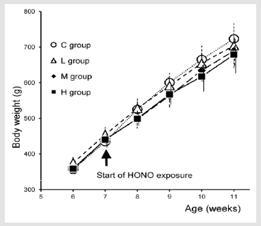
Figure 1: The transition of the body weight for each group. No significant differences were found by ANOVA.

Table 1: Concentrations of nitrogen oxides in the animal exposure chambers.
Although Guinea pigs were continuously exposed to HONO for 4 weeks, nitrogen oxides were sampled for 30 min per day for 5 days per week. aSingle measurement of collected HONO (ppm). bNOx analyzer data after removing HONO from sampled air (ppm).
Effects of HONO Exposure on the Bronchial Regions Under Light Microscopy with HE
Figure 2a shows the main bronchus near the middle of the rightlung middle lobe for each group (Figure 2). The bronchoconstriction varied by individual, and the comparison between groups was not clear. The boundary of the smooth muscle layer in the bronchial connective tissue was clear in each group. The average thickness of the bronchial smooth muscle layer near the middle of the rightlung middle lobe is shown in Table 2. Although a tendency for a thickening of bronchial smooth muscle with HONO exposure was observed, there was no significant difference. (Table 2) Figure 2b shows the magnified images of Figure 2a. There were no inflammatory alterations in any group. However, H group showed a tendency for hyperplasia and pseudostratification of bronchial epithelial cells. Figure 2c shows the alveolar duct regions of each group. Extension of bronchial epithelial cells in the alveolar duct regions was observed in a dose-dependent tendency for the thickened interstitium and the smooth muscle cells (arrows).
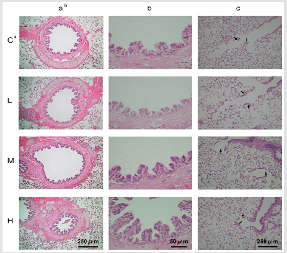
Figure 2: Effect of HONO exposure on the respiratory tract under light microscopy with HE. Stained with HE. a. Exposure groups: C; C group, L; L group, M; M group, H; H group. b. (a), (b) Main bronchus near the middle of right-lung middle lobe, (c) Alveolar duct regions.
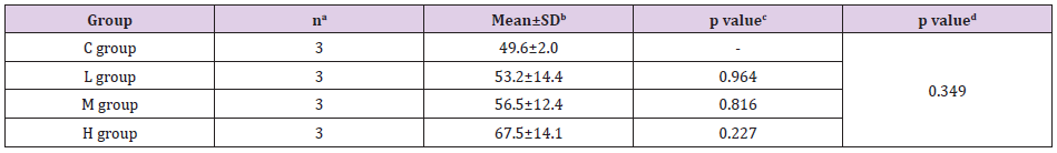
Table 2: Average thickness of the bronchial smooth muscle layer.
Dunnett’s multiple comparison (one-tailed) was applied for comparison with the control by ANOVA. aNumber of animals. bThickness of bronchial smooth muscle layer near the middle of right- lung middle lobe. cp value for comparison with C group. dp value for ANOVA.
Effects of HONO Exposure on Alveolar Regions Under Light Microscopy with EVG and TEM
Figure 3a shows the alveolar duct regions of each group (Figure 3). The hypertrophic alteration of the interstitium and the cells (arrows) was observed in the alveolar duct regions in H group. A flattened surface was observed in the alveolar duct regions in a dose-dependent tendency without fibrosis. Figure 3b shows the alveoli of each group. Pulmonary emphysema-like alterations were observed in the HONO-exposure groups with a dose-dependent tendency in the alveolar duct centriacinar regions. Morphometric quantification of the extent of emphysema-like alterations is shown in Table 3. The average Lm of each group was 39.8–59.4 mm. The Lm of H group was significantly longer than that of C group by Dunnett’s multiple comparison but not by ANOVA (Table 3). Figure 3c shows the interstitium of the alveolar duct regions of each group at 5000× magnification by TEM. Arrows indicate smooth muscle cells, which had rich mitochondria and intermediate microfilaments in L, M, and H groups, similar to Figure 2c observations. There were no edematous alterations in the alveolar regions of any group.
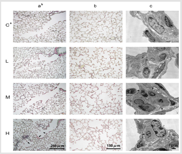
Figure 3: Effect of HONO exposure on the respiratory tract under light microscopy with EVG and TEM. a. Exposure groups: C; C group, L; L group, M; M group, H; H group. b. (a) The terminal bronchiole and alveolar duct regions, (b) The alveolar regions, (c) Interstitium of alveolar duct regions under TEM.
Table 2: Alveolar mean linear intercept in each group after 4 weeks of HONO exposure.
Dunnett’s multiple comparison was applied for comparison with the control by ANOVA. aNumber of animals. bAlveolar mean linear intercept. cp value for comparison with C group (*p < 0.05). dp value for ANOVA.
Effects of HONO Exposure on Histopathology under SEM
Figure 4a shows the bronchial lumen near the main bronchus of each group at 500× magnification by SEM. (Figure 4). The cilia of bronchial epithelial cells showed no damage in all HONOexposure group. The flattened surface of the bronchial epithelial cells was observed in the bronchial lumen in M and H groups, and this change had a dose-dependent tendency. Figure 4b illustrates the terminal bronchiole regions of each group at 100× and 200× magnifications by SEM. The proliferative alteration of the cells was observed in the alveolar duct regions in a dose-dependent manner. Figure 4c demonstrates the alveolar regions of each group at 200× magnification. Although the peripheral alveoli in C group were normally inflated, the peripheral alveoli in the HONOexposure groups (L, M, and H groups) were pulled in all directions and had small inflations in a dose-dependent manner. Although the peripheral alveoli of C group appeared to be expanding with low tension, the peripheral alveoli of the HONO-exposure groups (L, M, and H groups) appeared to be expanding with high tension. In contrast, the alveolar duct lumen of the HONO-exposure groups showed a dose-dependent expansion.
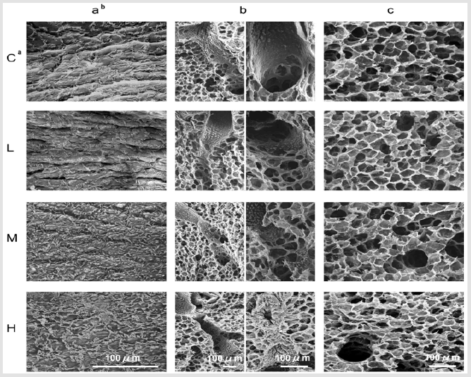
Figure 4: Effect of HONO exposure on the respiratory tracts under SEM. a. Exposure groups: C; C group, L; L group, M; M group, H; H group. b. (a) The bronchial lumen near the main bronchus, (b) The alveolar duct regions, (c) The alveolar regions.
Discussion
This was the first study to observe animal lungs from HONO exposure experiments with SEM and TEM. The SEM observations confirmed the pulmonary emphysema-like alterations in the centriacinar regions of alveolar ducts, which had been observed under light microscopy in our previous HONO exposure experiments. The SEM observations suggest that HONO exposure contracts the peripheral alveoli and expands the alveolar duct lumen. The pulmonary emphysema-like alterations were significant only in H group by Lm measurement, but the alterations were also observed in L and M groups using SEM. The TEM observations showed the presence of smooth muscle cells in the interstitium of alveolar duct regions in the HONO-exposure groups, and confirm that there were no injurious effects such as edematous alterations according to HONO exposure. In our previous HONO exposure experiments on animals, secondary products of NO2 and NO were sometimes present in the generated HONO, and the histopathological alterations that could be confirmed differed depending on the animal species.
This experiment had the lowest concentration of HONO in our HONO exposure experiments so far, secondary product of NO2 was not detected, and pulmonary emphysema-like alterations and a tendency for hyperplasia and pseudostratification of bronchial epithelial cells were observed. In previous HONO exposure experiments, the pulmonary emphysema-like alterations were observed in guinea pigs and rats, but not in mice. Although bronchial smooth muscle hypertrophy was identified in a previous HONO exposure experiment on rats, mice had few bronchial smooth muscle cells. Therefore we considered that HONO might induce architectural alterations in the smooth muscle cells. On the other hand, the hyperplasia of the terminal bronchial epithelial cells was remarkable after HONO exposure in mice, which used the highest concentration of HONO in our HONO exposure experiments so far. A tendency for hyperplasia and pseudostratification of bronchial epithelial cells was observed in this experiment.
The present results suggest that these alterations were due to HONO. In the present study, we observed pulmonary emphysema-like alterations in the centriacinar regions of alveolar ducts with a significant increase of Lm in H group, a tendency for hyperplasia and pseudostratification of bronchial epithelial cells, and the extension of bronchial epithelial cells and smooth muscle cells in alveolar duct regions; these results had a dose-dependent tendency. The present results suggest that the LOAEL of HONO is < 0.1 ppm by histopathological analyses. LOAEL is one of important grounds for Environmental Quality Standards (EQSs) for hazardous air pollutants. A review was reported for EQSs with guideline values for air pollutants including NO2. Although the review did not describe the LOAEL of NO2 from animal exposure experiments, it described that the epidemiological effects observed in residents showed a lower value than findings from animal exposure experiments and reports on human exposure.
It could be that the uncertainty factor (safety factor) was not necessary because the standards were based on data obtained from human subjects including those with high susceptibility [13]. The EQS for NO2 was set as ‘the daily average for hourly values shall be within the 0.04–0.06 ppm zone or below that zone’ in 1978 in Japan. The EQS for NO2 is similar to the LOAEL of HONO exposure experiments on animals. The present results suggest that HONO, which is detected as NO2, affects human health more than NO2 and that it is necessary to examine the involvement of HONO in the epidemiological studies of NO2. HONO existence in the atmosphere [14] and HONO contamination in NO2 measurements [1] are known factors. However, the EQS for NO2 and the measurement method of NO2 have not been revised since 1978, and the WHO Air quality guidelines described that it seems reasonable to retain an annual average limit value for NO2. The reason may be that previous studies on the health effects of HONO were not sufficient for the EQS for HONO.
Yoshida (1988) reported retrospectively the actual procedure for setting EQSs for classical pollutants including NO2 [15,16]. In the report, the findings from animal experiments, reports on human exposure, and epidemiological studies were collected, and the dose-effect/dose-response relationship was investigated; Generally, the most important data were the epidemiological findings. Among the studies on the health effects of HONO, our studies were the only animal exposure experiments [4-6], and there had been two studies on human inhalation [17,18] and three epidemiological studies, including our study [3,18,19]. The review of the EQS for NO2 states that scientists are in charge of proposing a guideline using scientific data, and the government is responsible for establishing its own standard. Therefore, we consider that the government should request scientists to provide opinions and new experiments on the HONO issue for revising the EQS for NO2 or establishing the EQS for HONO.
In conclusion, we found pulmonary emphysema-like alterations in the centriacinar regions of alveolar ducts, a tendency for hyperplasia and pseudostratification of bronchial epithelial cells, and the extension of the bronchial epithelial cells and smooth muscle cells in alveolar duct regions; these results had a dose-dependent tendency. In our previous HONO exposure experiments on animals, secondary products of NO2 and NO were sometimes present in the generated HONO. However, secondary product of NO2 was not detected in this HONO exposure experiment. Therefore, the present results suggest that these alterations were due to HONO.
Acknowledgments
The authors gratefully thank Hiroko Kyono, Ph.D., who guided us regarding the histopathological examination of specimens by transmission electron microscopy. This study is supported by a Grant-in-Aid for Scientific Research from JSPS KAKENHI Grant Number [JP20659098].
Declaration of Interest
The authors declare that there is no conflict of interest.
References
- Pitts JN Jr, Winer AM, Harris GW, Carter WP, Tuazon EC (1983) Trace nitrogenous species in urban atmospheres. Environ Health Perspect 52: 153-157.
- Harry M, Ten B, Henk S (1998) The dark decay of HONO in environmental (smog) chambers. Atmos Environ 32(2): 247-251.
- Ohyama M, Nakajima T, Minejima C, Azuma K, Itano Y, et al. (2019) Association between indoor nitrous acid, outdoor nitrogen dioxide, and asthma attacks: results of apilot study. Int J Environ Health Res 29(6): 632-642.
- Ohyama M, Oka K, Adachi S, Takenaka N (2010) Effects of nitrous acid exposure on pulmonary tissues in guinea pigs. Inhal Toxicol 22(11): 930-936.
- Ohyama M, Oka K, Adachi S, Takenaka N (2011) Histological effect of nitrous acid with secondary products of nitrogen dioxide and nitric oxide exposure on pulmonary tissue in mice. J Clin Toxicol 1(1): 1-4.
- Ohyama M, Horie I, Isohama Y, Azuma K, Adachi S, et al. (2018) Effects of nitrous acid exposure on baseline pulmonary resistance and Muc5ac in rats. Inhal Toxicol 30(4-5): 149-158.
- Shore S, Kobzik L, Long NC, et al. (1995) Increased airway responsiveness to inhaled methacholine in a rat model of chronic bronchitis. Am J Respir Crit Care Med 151(6): 1931-1938.
- Ohyama M, Oka K, Adachi S, Norimichi Takenaka (2013) Development of a gaseous nitrous acid generation system for animal exposure experiments. J Clinic Toxicol 3: 165.
- Koutrakis P, Wolfson JM, Slater JL, Brauer M, Spengler JD, et al. (1988) Evaluation of an annular denuder/filter pack system to collect acidic aerosols and gases. Environ Sci Technol 22(12): 1463-1468.
- Febo A, Perrino C, Cortiello M (1993) A denuder technique for the measurement of nitrous acid in urban atmospheres. Atmos Environ 27(11): 1721-1728.
- Dunnill MS (1962) Quantitative methods in the study of pulmonary pathology. Thorax 17(4): 320-328.
- Thurlbeck WM (1967) Internal surface area and other measurements in emphysema. Thorax 22(6): 483-496.
- Kawamoto T, Pham TT, Matsuda T, Oyama T, Tanaka M, et al. (2011) Historical review on development of environmental quality standards and guideline values for air pollutants in Japan. Int J Hyg Environ Health 214(4): 296-304.
- Platt U, Perner D, Harris GW, Winer AM, Pitts JN Jr (1980) Observations of nitrous acid in an urban atmosphere by differential optical absorption. Nature 285: 312-314.
- Yoshida K (1988) Ambient air quality standards. J Toxicol Sci 13: 262-264.
- Beckett WS, Russi MB, Haber AD, Rivkin RM, Sullivan JR, et al. (1995) Effect of nitrous acid on lung function in asthmatics: a chamber study. Environ Health Perspect 103(4): 372-375.
- Rasmussen TR, Brauer M, Kjaergaard S (1995) Effects of nitrous acid exposure on human mucous membranes. Am J Respir Crit Care Med 151(5): 1504-1511.
- van Strien RT, Gent JF, Belanger K, Triche E, Bracken MB, et al. (2004) Exposure to NO2 and nitrous acid and respiratory symptoms in the first year of life. Epidemiology 15(4): 471-478.
- Jarvis DL, Leaderer BP, Chinn S, Burney PG (2005) Indoor nitrous acid and respiratory symptoms and lung function in adults. Thorax 60(6): 474-479.
