 Case Report
Abstract
Two cases of unusual equine endometritis associated with Chaetomium sp are described in the article. The mares displayed infertility. The cytology revealed filamentous structures in both cases and the cultures yielded Chatomium sp which had never been described as an-endometritis agent. Chatomium sp growth usually in cellulose medium. The fungus, perhaps present on the stable floor, could colonize the uterus as an opportunist because of the failure in uterine defense mechanism.
Keywords: Mare; Fungal Endometritis; Chaetomium sp; Endometritis; Vaginal Discharge; lymphatic drainage
Introduction
Fungal endometritis refers to the inflammation of the uterine lining caused by the colonization of fungal organism. Because it creates an incorrect environment for an embryo, it is an important cause of infertility in the mare despite being relatively uncommon [1]. Actually, fungal growth is considered to be present in 1 to 5% of the cases. Yeast organisms most reported to colonize the equine reproductive tract include Candida spp and Aspergillus spp [2]. Clinical signs include subclinical infection to profuse vaginal discharge, moreover infertility is recurrent. The prognosis for fungal endometritis is guarded to poor [3].
Cases Details
Case 1
A 6-years-old Andalusian mare was referred to the Equine Reproduction Service (ERS), Veterinary Faculty of Barcelona in March 2004 because of infertility problems. The mare was born and bred in Spain, without previous foaling. The owner informed that the mare, when she was 3 years old, suffered an important vagino-cervical laceration as a consequence of a first mating, by a vigorous stallion with a large penis. A practitioner treated the laceration in field conditions; then the mare has been flushed, treated with intrauterine antibiotics and mated repeatedly, the following 3 years, but with no pregnancy result. In the ERS, the transrectal ultrasound examination showed an hyperechogenic cervix with two hypoechogenic areas that let guess a fibrotic cervix. Vaginoscopy reveals vagina and cervix lacerated, probably by the natural mating. Cytology sample was obtained by sterile uterine swab and revealed numerous polymorphonuclear neutrophils and unidentified filamentous structures. Endometrial swab culture evidenced the presence of Streptococus equi Zooepidemicus and fungi Chaetomium sp. The case was considered as a casual finding.
Case 2
A 10-years-old Swedich Warmblood mare was referred to the ERS in May 2016 because of infertility problems. The mare was born and bred in Sweden and had foaled 8 times. The last parturition was in 2013 and ended as a complicated dystocia. The owner informed that the mare has been flushed, treated with intrauterine antibiotics, inseminated and mated repeatedly without success. The external examination revealed a poor perineal conformation with much of the vulva positioned above the level of the pelvic floor (Figure 1) Vaginoscopy evidences an irregular and fibrotic cervix, probably because of the numerous parturitions and/or the dystocia (Figure 2) The transrectal ultrasound examination showed augmented intrauterine fluid during estrus. Cytology samples was obtained by uterine swab and no polymorphonuclear neutrophils were detected. Nevertheless, unidentified filamentous structures, similar to the first case, could be observed (Figure 3). No bacteria were evidenced by uterine samples culture. Samples, obtained by sterile swap, are plated on Sabouraud glucose plates, added with antibiotics, in order to avoid bacterial growth. The plates are incubated for seven to ten days at 28ºC. After this period of time fungal development in plaques is observed. In order to observe the fungal structure, it is necessary to prepare a fresh observation with lactophenol blue. The observation of the fungal structures, allows to deduce that it is a strain of the genus Chaetomium (Figure 4 and 5). On account to the difficulty to treat a fungal endometritis that, actually, was secondary to the poor perineal conformation and fibrotic state of the endometrium, and considering the correct welfare of the mare, it is decided to not treat the injurie.
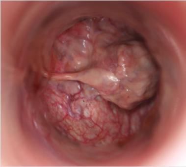
Figure 1: Fibrotic cervix in diestrus.
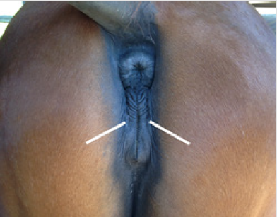
Figure 2: Poor perineal conformation. White lines are indicating the pelvis floor.
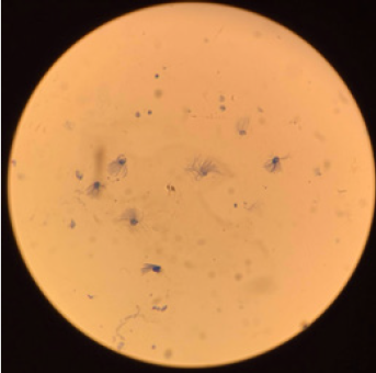
Figure 3: Endometrial cytology stained with Diff-Quik®.
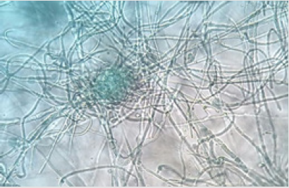
Figure 4: Initial development of the Chaetomium’s fruiting body (x400).
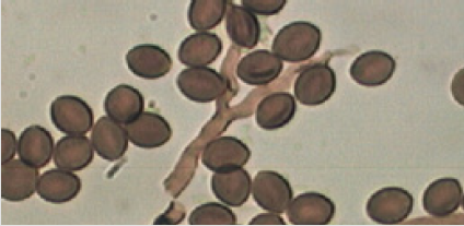
Figure 5: Chaetomium’s ascopores (x1000).
The histological evaluation of the uterine biopsy shows a luminal epithelium severely injured, loss of the stratum compactum, neutrophils and lymphocytes in small numbers. A severe periglandular fibrosis was observed, with fibrotic nests, lymphatic lacunae and cystic gland distension. Because of the highest level of pathological changes, the endometrial biopsy is classified as III grade (Kenney and Doig, 1986) according to the mare a low probability of carrying a foal to term (Figure 6).
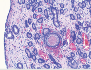
Figure 6: Histological evaluation of the uterine biopsy (x40).
Discussion
Mares predisposed to developing fungal endometritis may have abnormalities of the reproductive tract, generalized immunosuppression, and bacterial contamination in the first place. The use of intrauterine antimicrobials and repetitive flushes are also a cause of increase incidence of fungal endometritis, either through uterine contamination with fungal organisms, but especially by alteration of the normal bacterial flora of the reproductive tract. Once diagnosed, the treatment of fungal endometritis use to be unrewarding and recurrence of yeast infections is common [4]. Treatment involves physical removal of infectious organisms by uterine lavage, systemic or intrauterine administration of antifungal agents. Ideally, selection of an antifungal agent should be based on results of antifungigrams for each case of fungal endometritis and empirical treatment should be avoided [2]. As a chronic treatment, repair of abnormal conformation or damage in the reproductive anatomy should be performed. That includes surgical repair as the common Caslick’s procedure, perineal body reconstruction or cervical laceration repair [1].
To our best knowledge, this is the first report of Chatomium sp associated with equine endometritis. Chaetomium sp is a genus of filamentous fungi (Phylum Ascomycota, Class Sordariomycetes). Species of the ascomycete genus Chaetomium are important in the decomposition of plant and other cellulose-rich materials, and can be isolated easily from dung, plant debris and soil. The genus contains a number of species capable of growth at elevated temperatures as well as a few species that cause infections in vertebrates. Chaetomium globolusum is the most prevalent clinical species and mainly affect the nails and skin in humans. It also causes cerebral and systemic infections with high mortality rates [5]. Chaetomium globolusum is important to vertebrates’ health as a contaminant in indoor environments since it is known to produce mycotoxins [6].
Conclusion
The interest of this case report is that this fungus use to grow in cellulose medium, perhaps stable floor, and this has never been described as endometritis agent. Obviously, Chaetomium growth is considered to be secondary due to underlying factors impairing the local immunity. In fact, the first mare had an acute endometritis, with bacteria growth, and the second one had an-endometriosis. Defects in genital anatomy, lymphatic drainage, cervical function, excessive intrauterine manipulations facilitate the colonization of the uterus by opportunistic fungi. But, the real question is that if the fungus agent can survive in uterine medium, and if it would be contagious for human and/or others animals.
Acknowledgments
The authors declare no conflict of interest.
References
- Ferris RA (2012) Endometritis, Bacterial and Fungal. Clinical Veterinary Advisor pp. 166-168.
- Ferris RA (2015) Robinson’s Current Therapy in Equine Medicine. 7th Edition Science direct 689-691.
- Coutinho da Silva MA, Alvarenga MA (2011) Fungal endometritis Equine reproduction. 2nd ed John Wiley Sons 2: 2643-2651.
- Dascanio J, Ley W, Schweizer C (2000) How to diagnose and treat fungal endometritis. AAEP Proceedings 46: 316-318.
- Serena C, Montserrat Ortoneda, Josep Guarro (2003) In vitro activities of new antifungal agents against Chaetomium spp. And inoculum standardization. Antimicrobial Agents and Chenotherapy 47(10): 3161-3164.
- Pieckova E (2003) In vitro toxicity of indoor Chaetomium Kunze ex Fr. Ann Agric Environ Med 10(1): 9-14.
