 Research Article
Abstract
Introduction: The aim of this study was to evaluate the action of different endodontic irrigating solutions on the ultraestructural human dentin.
Materials and Methods: 1% sodium hypochlorite, 17% ethylendiaminetetraacetic acid, 1% calcium hydroxide, 0.2% chlorhexidine solutions and distilled water as control were used. 16 uniradicular human teeth recently extracted were washed and decoronated and the roots were longitudinally sectioned on the mesial and distal axes. The pulp was extracted and the root was symmetrically cut for comparison of the two half pieces. One of them was kept in contact with 1 ml irrigation solution for 30 minutes, while the other remained immersed in equal volume of sterile distilled water for the same time. Samples were processed according to the protocol for scanning electron microscope and analyzed at 1500X and 4000X. Surface residues, tubular holes open lumen, spaces between tubules, fibers, erosion, deposit of calcium of the middle dentin were considered. For the quantitative scanning electron microscopy analysis four parameters were observed: number of dentinal tubules by area in mm2, diameter tubular lumen, intertubular space, and relative density at 1500X. 20 measurements of each parameter for each middle third of the root dentin -control and treated- were performed, with Image Tool. Results were analyzed using the one-way ANOVA, Tukey tests and Pearson correlations.
Results: 1% sodium hypochlorite, 1% calcium hydroxide and 17% ethylendiaminetetraacetic acid produced significant qualitative and quantitative changes on the dentin surface and on the ultrastructure of the human dentin. 1% sodium hypochlorite and 1% calcium hydroxide showed solvent action. 1% calcium hydroxide produced alterations on organic component of the dentin. 17% ethylendiaminetetraacetic acid extracted calcium and left tissue fibers exposed. Significant changes were produced with 1% calcium hydroxide in diameter tubular, intertubular space and relative density, with 17% ethylendiaminetetraacetic acid in intertubular space and relative density. 0.2% chlorhexidinedid not eliminate the remnants of the surface nor modified the dentin ultrastructure.
Conclusion: 1% calcium hydroxide, 17% ethylendiaminetetraacetic acid and specially 1% sodium hypochlorite cleaned the remnants of the dentin surface and they also produced changes on the ultrastructure; 1% calcium hydroxide showed signs of alterations. 0.2% chlorhexidine was not effective in cleansing human dentin under the tested experimental conditions.
Keywords: Irrigation Solutions; Contact; Action; Human Dentin; Ultraestructure
Abbreviations: SEM: Scanning Electron Microscopy; EDTA: Ethylendiaminetetraacetic Acid; DW: Distilled Water
Introduction
Dentin is a composite material comprised of an inorganic component (50 vol %), an organic component (30 vol %), and water (20 vol %). The inorganic component contributes mainly to the stiffness of dentin. The main organic component of the dentin is type I collagen (90 %). These type I collagen fibrils are stabilizedby endogenous covalent intra and intermolecular crosslinks that contributesignificantly to the mechanical properties of the dentin. The structural integrity of the dentin, depends on the optimum balance between toughness and stiffness [1]. Different solutions were tested alone and alternatively [2] to clean the root canal during the endodontic treatment.There is no single irrigating solution with all beneficial properties. In the clinical treatment, different solutions are applied according to their properties [3] and certain concentrations are considered biocompatible and effective according to clinical researches [4,5]. Sodium hypochlorite (0.5%, 1%, 2.5% and 5.25% NaOCl); ethylendiaminetetraacetic acid (15% and 17%EDTA), chlorhexidine (0.12%, 0.2% and 2% CHx) solutions and calcium hydroxide (0.1%, 0.5%, 1% and 2.5% Ca(OH)2 suspensions in distilled water) are among the most used irrigating solutions during the mechanical preparation of the root canal.
Different effects on dentin of irrigating solutions during the endodontic treatment have been showed through scanning electron microscopy (SEM) studies [5,6].A study by scanning electron microscopy revealed increased efficiency of 17% EDTA respect to1% NaClO irrigants [6]. Too 1% Ca (OH)2 respect to 0.2% CHx [7].
Some authors showed that 2.5% NaOCl completely dissolved organic remains and that 17% EDTA removed the smear layer by chelating mineral detached mainly in the middle and cervical thirds of dentin [8-11], resulting in a cleaner surface and tubular lumen.A study by SEM with 17% EDTA, 42% citric acid and 5.25% NaOCl showed the erosive effect of these solutions in apical intraradicular dentin [12] chequear. The aim of this study was to evaluate the action of different irrigating solutions on the ultrastructure of the root dentin to facilitate the selection of the most suitable solution.
Material and Methods
Experimental Teeth and Solutions
16 freshly human maxillary incisors in intact state were extracted for orthodontic reasons in the clinic of the Faculty of Dentistry of the National University of Tucuman, from people of 17 to 33 years old were used. Teeth were externally washed with 1% NaOCl and distilled water (DW).1% NaOCl (Stanton, Buenos Aires, Argentina), 17% ethylendiaminetetraacetic acid (EDTA) (Biopack, Buenos Aires, Argentina), 1% Ca (OH)2 (Anedra Lab, Buenos Aires, Argentina) and 0.2 % chlorhexidine (CHx) (ICN BiomedicalsInc, Ohio, USA) were the irrigation solutions used. Steril distilled water (DW) was used as control.
Processing Substrates
Uniradicular upper incisors teeth without instrumentation were used. The crowns were removed and the root portions were sectioned with a high speed turbine cylindrical diamond stone with cooling along their long axis in the vestibule lingual direction under water spray coolant. The pulp was extracted and the two half roots were used for comparison. Half root of each tooth layed in 1 ml of each irrigation solution during thirty minutes; the other half was immersed in the same volume of DW (control) for the same time. We worked in quadruplicate.The half roots were carefully washed with DW. Then they were processed according to the SEM protocol (1°- glutaraldehyde fixation 2°- 100% ethanol and acetone de hydration; 3°- freeze dehydration up to the critical point of freezedrying; 4°- placing the roots on stubs; 5°- golden metallization). The specimens were mounted on aluminum cylinders 3 cm in diameter and gold- coated. They were observed using SEM (JSM-35CF; Scanning microscope, JEOL, Tokyo, Japan) at 20X magnification to select the area of observation in the middle third of the root dentin; a line was drawn on the major root axis in the middle of the canal, and a perpendicular line was drawn at 2 mm from the apex. Photomicrographs was standardized at 1500X y 4000X of the dentin wall of the canal and observed by two evaluators. Qualitative Analysis. For the qualitative study, the morphological characteristics of the media dentin surface: surface residue (R), tubular hole open lumen (T), space between tubules (S), fibers (F), erosion (X), calcium deposit (Ca) were observed with SEM.
Quantitative Analysis
In the middle third of human dentin four parameters according to the measurement micrometric scale were quantified with an image analyzer as measuring scale Image Tool, sensibility 1/100 μm.T/A: number of dentin tubules per area in mm2 (3.5 mm = 1μm; micrographic area = 50 x 33 μm) (13) TD: the biggest tubule hole open lumen [14]. ITS: intertubular space equivalent to the space between tubules in the area [15]. RD: relative density related to the relation between the tubular area and the area of the micrograph/ 100X [14-16] to 1500x. 20 measurements per parameter were made for each root (the treated and the controlones).
Statistical Analysis
The variables were analyzed with Anova factorial test. Levenne test was used to determine the homogeneity of the variances of T/A, TD, ITS and RD variables. The Tukey test was applied to compare results of different solutions with an alpha error α = 1%. The parametric correlation of Pearson was used to compare TD, ITS and RD variables. The analysis between the treated and control roots was performed with the T test for independent samples previously analyzing the normality of the variables (Kolgomorov– Smirnov Test) with an alpha error α = 1%. Data were analyzed with the SPPSS statistical software 11.0 (Inc, Il USA).
Result
Qualitative Analysis
The control dentin showed many cleared dentin tubules (Figure 1A), and others with their holes partially filled with surface residues which were also observed in the intertubular spaces. At 4000X collagens fibers were observed in the matrix and in the tubular holes. After the use of 1% NaOCl the surface and holes of the tubules could be observed without residues. The middle third dentin structure showed similar aspect than of the control group; the intertubular spaces kept their aspect and distribution, although a principle of erosion could be seen and the collagen fibers were less visible (Figure 1B), 4000X). After the use of 17% EDTA (Figures 2A & 2B) the dentin surface was partially cleared of residues, with more clear tubular holes open lumen, intertubular space with signs of erosion and more visible collagen fibers with respect to the control group (Figure 2A).
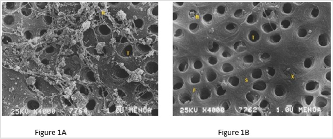
Figure 1: Figure 1A: Scanning electron micrography of root dentin Control (4000X); bar = 1.0 μm. Figure 1B: Scanning electron micrograph of root dentin 1% NaOCl (4000X); bar = 1.0 μm. R: Surface Residues, T: Tubular Hole Open Lumen, S: Space Between Tubules, F: Fibers, X: Erosion.
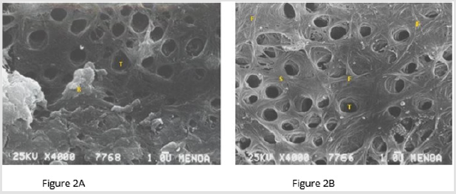
Figure 2: Figure 2A: Scanning electron micrograph of root dentin Control (4000X); bar = 1.0 μm. Figure 2B: Scanning electron micrograph of root dentin 17% EDTA 4000X); bar = 1.0 μm. R: Surface Residues, T: Tubular Hole Open Lumen, S: Space Between Tubules, F: Fibers, X: Erosion.
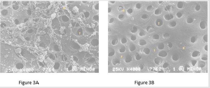
Figure 3: Figure 3A: Scanning electron micrograph of root dentin Control (4000X); bar = 1.0 μm. Figure 3B: Scanning electron micrograph of root dentin 1% Ca (OH)2 (4000X); bar = 1.0 μm. R: Surface Residues, T: Tubular Hole Open Lumen, S: Space Between Tubules, F: Fibers, X: Erosion.
In Figure 3A were observed dentin tubules cleared from residues, with reduced intertubular spaces, signs of erosion (X) and few but clearly visible collagen fibers after the use of 1% Ca(OH)2; bigger tubular holes partially cleared and with few residues with respect to the control group (Figure 3A). With DW (Figure 4-right) and 0.2% CHx (Figure 4A) the root dentin showed surface and tubules of similar aspect and distribution (4000X); collagen fibers were evident in both (F) as well as cleared tubule holes (T) with similar diameters and some of them with their holes partially filled with surface residues (R).
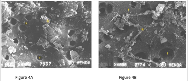
Figure 4: Figure 4A: Scanning electron micrograph of root dentin Control (4000X); bar = 1.0 μm. Figure 4B: Scanning electron micrograph of root dentin 0.2% CHx (4000X); bar = 1.0 μm. R: Surface Residues, T: Tubular Hole Open lumen, S: Space Between Tubules, F: Fibers, X: Erosion.
Quantitative Analysis
All quantized values in the middle third of human root dentin Control showed inactivity presenting distilled water (Table 1). When comparing the values obtained between each parameter and its controls; the following data is obtained: 1% NaClO: T / A (p <0.001) and DT (p <0.001) decrease; EIT (p <0.005) and DR (p <0.001) increased (Table 2A); 17% EDTA: DT (p <0.005) and EIT (p <0.001) increase; DR (p <0.001) decreases (Table 2B); 1% Ca (OH)2: DT (p <0.005) and DR (p <0.001) increased with treatment (Table 2C); 0.2% chlorhexidine: are not significantly altered the parameters compared to controls (p~ 0.01) (Table 2D).
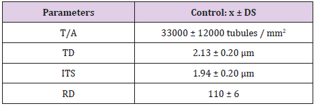
Table 1: Mean and Standard Deviation of parameters in Control dentin (n=20).
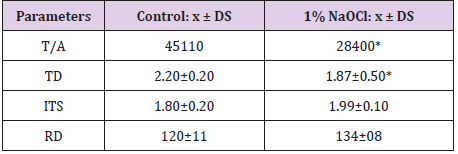
Table 2A: Mean and Standard Deviation of parameters in Control and 1% NaOCl dentin.
Note: *Significant difference from the control (p <0.001).
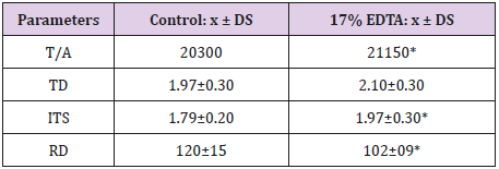
Table 2B: Mean and Standard Deviation of parameters in Control and 17% EDTA dentin.
Note: *Significant difference from the control (p <0.001).
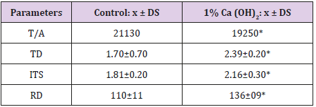
Table 2C: Mean and Standard Deviation of parameters in Control and 1% Ca (OH)2 dentin.
Note: *Significant difference from the control (p <0.001).
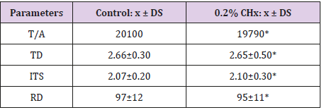
Table 2D: Mean and Standard Deviation of parameters in Control and 0.2% CHx dentin.
Note: *The differences are not significant (p≥0.05).
Statistical Analysis
High dispersion of T/A was observed in control (p <0.01) (Figure 5); in RD low dispersion was observed for both groups (p <0.001). When comparing among solutions, TD showed high dispersion (Figure 6) specially with Ca (OH)2; TD increased (p <0.001) and showed a negative difference. Low dispersion was observed in RD with all the solutions with respect to CHx; RD increased with Ca (OH)2 (p <0.001) and lowered with NaOCl (p <0.01). ITS showed high dispersion (Figure 7) especially in root dentin with Ca (OH)2 (p <0.001). T/A and TD decreased (p <0.001) where as ITS and RD increased with 1% NaOCl respect to control (Figure 8). RD decreased with 17% EDTA (p <0.001) (Figure 9). TD, RD and ITS increased with 1% Ca (OH)2 (p <0.001) (Figure 10). No significant differences were observed respect to control with 0.2% CHx (p ~ 0.01).
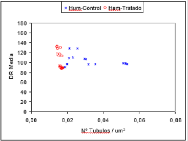
Figure 5: Comparative distribution area and tubules in dentin relative density control and contacted with the irrigation solutions.
Note: RD: Average Relative Density; N°T/μm2: Tubules/ Area; Hum-Control: Human Control; Hum-Tratado: Human Treated
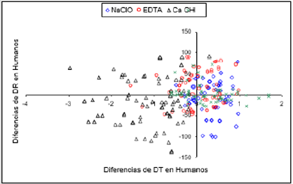
Figure 6: Distribution of differences of tubular diameter and relative density between control dentin and dentin which contacted the irrigation solutions.
Note: RD: Relative Density of Humans; TD: Tubular Diameter of Humans; NaOCl: 1% Sodium Hypochlorite; EDTA: 17% Ethylenediaminetetraacetic Acid; Ca (OH)2: 1% calcium hydroxide; CH: 0.2% chlorhexidine
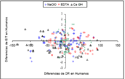
Figure 7: Distribution of differences of relative density and human dentin intertubular space between control and contacted with the irrigation solutions.
Note: RD: Relative Density of Humans; ITS: Intratubular Spaces of Humans; NaOCl: 1% Sodium Hypochlorite; EDTA: 17% Ethylenediaminetetraacetic Acid; Ca (OH)2: 1% Calcium Hydroxide; CH: 0.2% Chlorhexidine
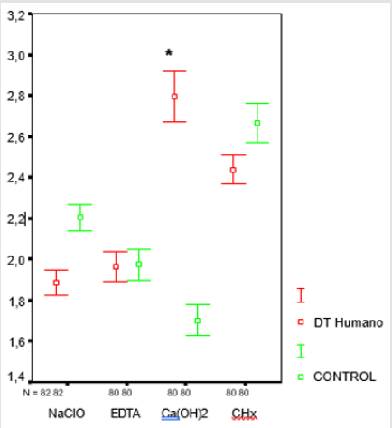
Figure 8: Media and confidence interval (CI) of the TD variable human dentin contact control and irrigation solutions.
Note: CI: Media and Confidence Interval of Control and
Treated Humans; DT: Human Tubular Diameter; Control:
Distilled Water; NaOCl: 1% Sodium Hypochlorite; EDTA:
17% Ethylenediaminetetraacetic Acid; Ca (OH)2: 1% Calcium
Hydroxide; CHx: 0.2% Chlorhexidine
*Significant difference from control (p <0.001)
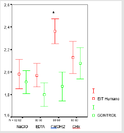
Figure 9: Media and confidence interval (CI) of the ITS variable human dentin contact control and irrigation solutions.
Note: CI: Media and Confidence Interval of Control and
Treated Humans; ITS: Intratubular Spaces Of Human;
Control: Distilled Water; NaOCl: 1% Sodium Hypochlorite;
EDTA: 17% Ethylenediaminetetraacetic Acid; Ca
(OH)2: 1% Calcium Hydroxide; CHx: 0.2% Chlorhexidine
*Significant difference from control (p <0.001)
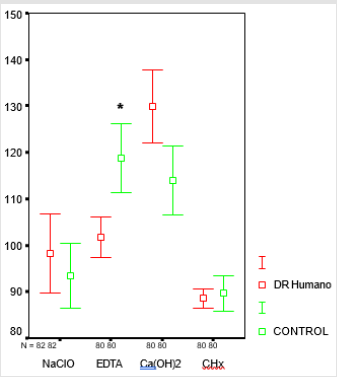
Figure 10: Media and confidence interval (CI) of the RD variable human dentin contact control and irrigation solutions.
Note: CI: Media and Confidence Interval of Control and
Treated Humans; RD: Relative Density Of Human; Control:
Distilled Water; NaOCl: 1% Sodium Hypochlorite;
EDTA: 17% Ethylenediaminetetraacetic Acid; Ca (OH)2:
1% Calcium Hydroxide; CHx: 0.2% Chlorhexidine
*Significant difference from control (p <0.001)
Discussion
This study shows the changes that endodontic solutions produced both on the structure and the ultrastructure of the root dentin: 1% NaOCl and 1% Ca (OH)2 removed surface residues and cleared the dentinal tubule holes. The solvent and proteolytic action of NaOCl affected the intertubular collagen according to Agee et al. [17] and Miori Pascon et al. [18]. Ca (OH)2 partially dissolved surface residues and showed signs of alteration in the ultraestructure. The intertubular spaces were damaged and peritubular dentin absence was observed.17% EDTA did not affect surface remnants although the intertubular dentin fibers became more evident because of calcium extraction. The organic matrix of the dentin would act as a limiting factor for the demineralizing effect of the solution. Little erosion could be observed in different areas. O´Conell et al. [19] showed that chelating action increased on previously treated dentin with 2.5% NaOCl which produced protein denaturalization that would facilitate dentin demineralization.The lonely application of 17% EDTA according to Mota Moreira et al [20] produced demineralization on bovine dentin whereas 5.25% NaOCl dissolved matrix collagen. When alternated, the solutions produced dissolution and loss of tissues. Long-lasting treatments with the chelating agent also produced dentin weakening because of micro-hardness decrease and organic component loss [21].
The alternated use of different solutions was frequent because none of them isolated met all the requirements of the treatment for the right cleansing of the canal and the bonding of sealants to the dentin. Nevertheless it is important to consider the erosion that it could produce on the dentin tissue [22] and its influence on micro- hardness and posterior resistance [21,23]. Agee et al. [17] mentioned that when 17% EDTA removes the mineral component, it exposes the collagen fibers and tissue becomes more vulnerable to proteolytic degradation. Calcium and phosphorous give more strength and hardness to the matrix and collagen fibers give resistance and flexibility to tissue.The dentin organic componentcollagen-, gives resistance to fracture, whereas enamel, with a high mineral content, is more rigid and fragile.
The solution, its concentration, the alternate use of different solutions and their interactions, the contact time between dentin and the solutions and the areas of the root to be treated should be considered. To standardize the study, the most representative middle third of dentin was chosen. % 0.2 CHx does not modify neither the surface nor the dentin structure [24]. In re-treatment of roots with high amount of gram positive bacteria CHx was successfully applied as a final rinse, after drying the NaOCl remnantin the root canal.The statistical study of human substrates controls were widely dispersed; a heterogeneous distribution T/A dentin was evident. TD, RD and ITS had lower dispersion and the like, indicating a selection of homogeneous substrates young age.
After contacting the solutions 1% NaOCl significantly diminished T/A and TD and increased ITS and RD. 17% EDTA significantly diminished RD and slightly increased the rest of the parameters. 1% Ca (OH)2 significantly increased TD, ITS and RD.0.2% CHx did not produce changes in the studied parameters during 30 minutes contact. Soares et al. [25] showed through SEM and a polarized light microscope that 0.12% CHx in contact with human dentin does not affect its morphological and mechanical properties because it does not produce collagen degradation. Other studies showed that its alternate use with 2.5% and 5% NaOCl affects the organic component and mechanical resistance of human dentin [11,24,26] which rises doubt about its biocompatibility. Ergucu et al. [27] stated that Na- OCl at low concentrations was biocompatible and adequate for the human root canal and dentinal tubules cleaning.
The statistical study of the parameters revealed low dispersion which a homogeneous response to the solutions action except to Ca (OH)2 1% high dispersion in TD and ITS. The negative half show significant increase in value after contact with the solution.1% Ca (OH)2 raised ITS, RD and specially TD. 1% NaOCl diminished T/A and TD. 17% EDTA diminished RD, increased ITS and TD. 0.2% CHx did not significantly modify the measured parameters with respect to the controls.The results of this study demonstrated the biocompatibility with dentin of 0.2% CHx, although it was not efficient in the dentin surface and canals cleaning under the experimental conditions. The other solutions modified the ultrastructure of dentin, specially 1%Ca (OH)2. For an optimal cleaning of the canals inhuman root dentin, 1% NaClO and 17% EDTA would be biocompatible and efficient agents used under low concentrations and at less contact time.
Conclusion
1% NaOCl and 1% Ca (OH)2 endodontic solutions dissolved surface remnants, affected fibers of dentin and opened the lumen of tubular holes after 30 minutes contact with dentin. 1% NaOCl showed effective solvent action on surface remnants and 1% Ca (HO)2 showed a greater action on the ultrastructure of dentin. It also produced significant changes on the intertubular and peritubular dentin surface.17% EDTA did not eliminate remnants of the dentin surface, extracted calcium, and exposed organic component and fibers. Quantified parameters showed significant changes with 1% Ca (OH)2 in TD, ITS and RD, and with 17% EDTA in ITS and RD. 0.2% CHx was inefficient in its cleaning under the tested experimental conditions.
Acknowledgement
This study was partly funded by the Odontology Faculty and the Science, Art and Technological Innovation Council at National University of Tucuman.
Conflict of Interest
No conflict of interest.
References
- Shrestha A, Friedman S, Kishen A (2011) Photodynamicaly Crosslinked and chitosan-incorporated dentin collagen. J Dent Res 90(11): 1346-1351.
- Mc Comb D, Smith D, Beagrie G (1976) The results of in vivo endodontic chemomechanical instrumentation. A scanning electron microscopic study. J Brit Endod Soc 9: 11-18.
- Dogan H, Calt S (2001) Effects of chelating agents and sodium hypochlorite on mineral content of root dentin. J Endod 27: 578-580.
- Hauman C, Love R (2003) Biocompatibility of dental materials used in contemporary endodontic therapy: a review. Part 1. Intracanal drugs and substances. Int Endod J 36: 75-85.
- Carvalho A, Camargo C, Valera M, Camargo S, Gasparoto Mancini M (2008) Smear layer removal by auxiliary chemical substances in biomechanical preparation: A scanning electron microscope study. J Endod 34: 1396-1400.
- Vasconcelos B, Luna Cruz S, De Deus G, Moraes I, Maniglia Ferreira C, et al. (2007)Cleaning ability of chlorhexidine gel and sodium hypochlorite associated or not with EDTA as root canal irrigants: A scanning electron microscopy study. J Appl Oral Sci 15(5): 387-391.
- Barbin L, Saquy P, Costa Guedes D, Sousa Neto M, Estrela C, et al. (2008) Determination of para- chloroaniline and reactive oxygen species in chlorhexidine and chlorhexidine associated with calcium hydroxide. J Endod 34(12): 1508-1514.
- Verdelis K, Eliades G, Oviir T, Margelos J (1999) Effect of chelating agents on the molecular composition and extent of decalcification at cervical, middle and apical root dentin locations. Endod Dent Traumatol 15(4): 164-170.
- De la Casa M, Raiden G (2005) A scanning electron microscopy evaluation of different root canal irrigating solutions. Acta Odontol Latinoam 18(2): 57-61.
- Zehnder M (2006) Root Canal Irrigants. J Endod 20: 1-10.
- Sirtes G, Waltimo T, Schaetzle M, Zehnder M (2005) The effects of temperature on sodium hypochlorite short- term stability, pulp dissolution capacity, and antimicrobial efficacy. J Endod 31(9): 669-672.
- Mancini M, Armellin E, Casaglia A, Cerroni L, Cianconi L (2009) A comparative study of smear layer removal and erosion in apical intraradicular dentin with three irrigating solutions: A scanning electron microscopy evaluation. J Endod 35(6): 900-903.
- Dutra Correa M, Anauate Netto C, Arana Chavez V (2007) Density and diameter of dentinal tubules in etched and non-etched bovine dentin examined by scanning electron microscopy. Arch Oral Biol 52(9): 850-855.
- Schilke R, Lisson JA, Baub O, Geurtsen W (2000) Comparison of the number and diameter of dentinal tubules in human and bovine dentin by scanning electron microscope investigation. Arch Oral Biol 45(4): 355-361.
- Huang T, Gulabivala Y, Ng L (2008) A bio-molecular film ex-vivo model to evaluate the influence of canal dimensions and irrigation variables on the efficacy of irrigation. Int Endod J 41(1): 60-71.
- Zeman L (2011) Comportamiento de los ionómeros vítreos y los composites en el medio bucal de pacientes bulí Doctoral thesis. UNT. Tucumán, Argentina.
- Agee K, Zhang Y, Pashley D (2000) Effects of acids and additives on the susceptibility of human dentin to denaturation. J Oral Rehabil27(2): 136-141.
- Miori Pascon F, Kantovitz K, Sacramento P, Nobre dos Santos M, Puppin Rontani R (2009) Effect of sodium hypochlorite on dentin mechanical properties. Review. J Dent 37(12): 903-908.
- O´Connell M, Morgan L, Beeler W, Baumgartner J (2000) A comparative study of smear layer removal using different salts of EDTA. J Endod 26(12): 739-743.
- Mota Moreira D, Almeida J, Randi Ferraz C, Figueiredo B, Reres Line S, et al. (2009) Structural analysis of bovine root dentin after use of different endodontic auxiliary chemical substances. J Endod 35(7): 1023-1027.
- Kamakshi G (2014) Relation between calcium loss and its effect on microhardness of root canal dentin following treatment with 17% EDTA at different time intervals: An ex vivo study. J Int Med and Dent 1(2):75- 85.
- Mancini M, Armellin E, Casaglia A, Cerroni L, Cianconi L (2009) A comparative study of smear layer removal and erosion in apical intraradicular dentin with three irrigating solutions: A scanning electron microscopy evaluation. J Endod 35(6): 900-903.
- Cem Sayim T, Serper A, Cebreli Z, Kalayci S (2007) Calcium loss from root canal dentin following EDTA, EGTA, EDTAC, and tetracycline-HCl treatment with or without subsequent NaOCl irrigation. J Endod 33(5): 581-584.
- Tanomaru Filho M, Leonardo M, Silva L, Aníbal F, Faccioli L (2002) Inflammatory response to different endodontic irrigation solutions. Int Endod J 35(9): 735-739.
- Soares C, Neiva R, Soares P, Dechichi P, Novais V, et al. (2011) Effects of chlorhexidine and fluoride on irradiated enamel and dentin. J Dent Res 90(5): 659-664.
- Marending M, Paque F, Fischer J, Zehnder M (2007) Impact of irrigant sequence on mechanical properties of human root dentin. J Endod 33(11): 1325-1328.
- Ergucu Z, Hiller K, Schmalz G (2005) Influence of dentin on the effectiveness of antibacterial agent. J Endod 31: 124-129.
