 Research Article
Abstract
Breast cancer is a heterogeneous disease that represents a major public health problem, it is the most common cancer and leading cause of cancer deaths among Nigerian women, a condition that may be predicated upon by inadequate knowledge about fundamental regimen necessary for cancer prevention, early detection and unavailability of necessary health requirement. Immunohistochemistry is now a standard methodology in pathology laboratories used in providing substantial information with regard to diagnosis, therapeutic prediction and prognosis of breast cancer. Little is known about the biology, molecular profile and optimal treatment of African Nigerian breast cancer. This article discusses the immunohistochemical and prognostic markers of breast cancer in Nigeria. Literature was reviewed mainly through Public Medline search with the terms “immunohistochemical, prognosis markers of breast cancer in Nigeria”, hard copies of journal pages and conference paper on prognostic markers of breast cancer in Nigeria. There are many markers that characterizes breast cancer and provide information about choice of therapy and prognosis, but as a developing country, the need for effective enlightenment and screening programs at an earlier age is required to boost the fight against the dreaded disease called breast cancer ravaging our women.
Keywords: Cancer; Breast Cancer; Prognostic Markers; Immunohistochemical Markers
Abbreviations: NHL: Non-Hodgkin’s lymphoma; ER: Estrogen Receptor; PR: Progesterone Receptor ER-LBA: Radio-Ligand Binding Assay
Introduction
Nigeria is the most populous black country on earth with over 152 million people, it is located in the African continent at the western coast. It features 36 states with four main geographic region which includes the coast, the forest belt, the savannah, and the Sahel [1]. Cancer is an abnormal growth of cells in the body. Clinically, it is defined as an enormous number of complex diseases that behave contrarily and proliferate into surrounding cells [2]. Cancer is a global public health burden affecting all categories of people, it is among the three leading causes of death in developing countries and the second most common cause of death in developed countries [3]. The fast spread of this disease might be attributed to unawareness of its early symptoms which can be treated at its early stage when detected. Cancer proliferation and metastasis nature made it a dreadful disease, this has resulted to its fast and quick spread in the body [4]. Cell proliferation is the continuous division of cells into multiple units without control. Metastasis is a process that allows spreading of the cells to other parts of the body. The interesting and painful aspect of this dreaded disease is its ability to pool both cell proliferation and metastatic making it more hazardous [2].
WHO [3] reported ‘24.6 million people globally lives with cancer from which approximately 7 million death occur with an estimated 11 million new cases every year’. Globally, cancer accounts for 12.5% of all deaths and if there is a tendency that by 2020, 16 million new cases will be diagnosed yearly and developing countries will account for 70% of the cases [3]. Popoola et al. [4] reported ‘650,000 people of estimated 965million Africans are diagnosed of cancer annually and women are at a higher risk. Common cancers in Nigeria in ascending order of incidence include: Non- Hodgkin’s lymphoma (NHL), colorectal, liver, prostate, cervical and breast cancer [5]. Breast and cervical cancer are the most common cancer in females [6].
Breast cancer is the most common malignant tumor in women although its mortality is declining gradually due to advanced diagnostic and therapeutic methods in developed countries. Nevertheless, this cancer-related mortality remains very high in the developing countries because of late diagnosis associated with a delay in treatment [7]. It is a heterogeneous disease with various prognostic and therapeutic implications [8].The incidence and clinical outcomes of breast cancer differ across various populations, age and racial groups, with higher incidence rates reported in developed countries, In many developing countries, however, its incidence has recently been found to be on the increase and in Nigeria, breast cancer has overtaken cervical cancer as the leading cause of cancer mortality in women [4]. According to Thull and Vogel (2004), About 5% to 10% of all breast cancer cases are estimated to be strongly hereditary. BRCA1 and BRCA2 are the most commonly mutated genes of cancer, but additional genes associated with the hereditary breast cancer are appearing [9]. Other inherited cancer genes that predispose to breast cancer include TP53 mutations in Li-Fraumeni syndrome, STK11 mutation in Peutz-Jeghers syndrome and P10 mutations in Cowen syndrome [10]. Somatic (not-inherited) mutations in the PIK3CA oncogene are common in human breast cancer; mutations are observed in 20% to 40% of cases [11].
The risk factors for breast cancer includes: increasing age, family history, young age at menarche, late menopause, overweight after menopause, null parity or late age at first birth, breast density, long-term use of combined estrogen progestin hormone therapy after menopause, certain types of benign breast diseases and alcohol consumption [12]. However, only a small number of women that develop breast cancer carry the above-mentioned risk factors [13]. Tumor size, lymph node involvement, and distant metastasis are factors traditionally known to predict breast cancer behavior and management although the clear picture of the behavioral pattern of breast cancer still remains unclear [14].
Breast cancer has a high mortality rate among African women, it is usually characterized by late presentation mostly at an advanced stage when compared to Caucasian American and European women [4]. and this might be attributed to inadequate access to good health care system. In the USA, the 5-year relative survival of African American women diagnosed with breast cancer is 77.7% [15]. Data has shown that Black women have a relatively higher frequency of biologically aggressive breast cancer that are hormone receptor negative [16]. There is a low survival rates in lowincome countries and can be attributed to socio-economic status, ineffective early detection programs, poor access to timely health care services and the lack of adequate diagnostic and treatment facilities, the inherent biology of the disease itself may play a role in the adverse outcome [4].
According to Khaitan [17] Immunohistochemistry is a technique used in identification of cellular or tissue constituents (antigens) by means of antigen-antibody interactions or by use of secondary labelling method. The use of immunohistochemistry to further characterize breast cancer globally has introduced a new dimension to the knowledge of breast cancer, it can no longer be regarded as a single entity and morphological features alone cannot completely predict the behavior of breast cancer [18] and also the use of molecular markers along with the traditional biological markers is been used to produce a better index for the prediction of breast cancer risk, assessment of prognosis in breast cancer prone environment and selection of appropriate treatment, for example, trastuzumab (Herceptin) a monoclonal antibody against ERBB2 has shown clinical benefit in 15-20% of ERBB2 positive breast cancer patients [19].Immunohistochemical markers currently in routine diagnostic use in most countries are estrogen receptor (ERα), progesterone receptor (PR) and Her2 [20]. According to the National Coordinating Group for Breast Screening Pathology (2008), the markers determine which tumors are likely to respond to hormonal therapy and Herceptin treatment (Figure 1).
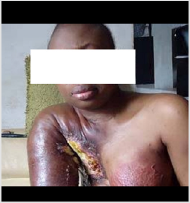
Figure 1: A Nigerian woman with breast cancer.
Source: Nigerian Vanguard Online news outlet.
Immunohistochemical & Prognostic Markers of Breast Cancer in Nigeria
Classical Prognostic Markers
Stage of The Disease
According to Heiden and DeBerardinis [21] ‘The breast cancer stage of an affected individual has a direct impact on patient outcome. The stage is defined by pTNM classification which comprises of tumour size, histological sections measurements (pT), extent of axillary nodal involvement, number of involved lymph nodes investigated histologically (pN), the extent of distant metastases, verified histologically or cytologically [4]. According to Leena [22], there is no particular treaty on the critical size of tumour cell clusters that should be regarded as metastes
Tumour Grade
Histologic grade is another prognostic factor for breast cancer, it was introduced by Bloom and Richardson in the early 1950s and this grading has been validated [24]. Tumour grade consist of the aptitude of cancer cells to form glandular structures, their nuclear morphology and mitotic counts [22]. According to Leena [22] tumour grade, nuclear and mitotic counts are often considered as separate, independent prognostic markers especially when analysed by morphometric methods. The classical prognostic markers are well established and validated. They form the cornerstone of breast cancer diagnostics, and all other prognosis indicators are tested against them, and it is not an exemption in Nigeria [23]. A study by Jennings [24] on clinicopathological features and molecular markers of breast cancer in Jos, Nigeria showed and concluded that 70.6% of breast cancer Tumor among Nigerians is high grade.
Cancer Type
Breast cancer is typed according to its morphology and named after the presumed cellular origin in the terminal duct-lobular unit [22]. Breast cancer is largely divided into ductal carcinomas comprising 10-30% of breast cancers, as reported by Jennings [24], small cell ductal carcinoma occurs in special subtypes, including tubular and certain papillary carcinomas and large cell ductal breast carcinoma grows in several patterns, metaplastic, medullary and infiltrating micropapilary carcinoma. According to Silverstein et al [25], A ductal carcinoma frequently evokes an inflammatory reaction which is rarely seen in lobular carcinoma and Lobular breast carcinomas are characterized by small cells with a scanty cytoplasm. Majority of cases retrieved by [24] from hospitals in Nigeria showed that majority of the cases were invasive ductal carcinoma (90.7%).
Other Prognostic Markers
Several global studies have presented persuasive evidence to support prognostic importance of the recognition of tumor cells invading lymphatic and blood vessels, However, its application is seriously disadvantaged by inter-observer and intra-observer differences in interpretation. A more uniform and objective approach, such as the use of immunohistochemical techniques in identifying endothelial linings, may be helpful in overcoming these obstacles [22].
Patient-Related Prognostic Markers
Age of Patients
Age has an impact in the basic physiological processes of the body [26]. Hormones that produces tissues and female reproductive organs are specifically affected. Although, physiological proliferation of the epithelia slows down with age, the cumulated damage to the genome of epithelial cells increases with time and this has an impact on breast cancer in a twofold manner [22]. Cancer is more prevalent in postmenopausal patients and it is usually more rapidly progressive. Clinical, in young patients, the tumour size is larger whichsuggests higher stromal activity [26]. Different variables have been found to correlate significantly with shortened recurrencefree survival in premenopausal women: the age, large tumour size, high number of metastatic lymph nodes in the axilla, high histological grade, and negative ER and PR status of the tumour. In multivariate analyses, young age is the most important adverse factor in premenopausal patients, followed by tumour size and histological grade, whereas PR status is of borderline significance [22]. A study was done by Ikpat & Collan [26] to access the effect of age as a prognostic factor, he used univariate and multivariate Cox’s regression on a retrospective study of invasive breast cancer seen in Calabar over a seventeen-year period in University of Calabar Teaching Hospital, Calabar, Nigeria. He concluded that Patients less than 40 years accounted for 39.8% of the total number of patients with infiltrating breast carcinoma and that there was a positive association between age and tumour.
Molecular Prognosis Markers of Breast Cancer
Genetics of Breast Cancer
Malignant cells are described by unstable genome characteristics, making their behaviour erratic [27]. In the classical mouse melanoma metastasizing experiments, Fidler and Nicholson in 1973 showed that cells with different surface properties had a different propensity for metastasizing into selected organs. They also found that primary tumors contained sub-clones of cells having different cell surface properties. They thus proposed that this might be at least partially caused by post-transcriptional heterogeneity, due to different glycosylation of the same surface molecules.
Flow cytometric DNA analysis of breast cancer yields information on the DNA content of single cells, knowledge of the DNA synthesis phase fraction gives important prognostic information. The National Institute for Medical Research, Yaba, Lagos, Nigeria has laboratories where such analysis is done in Nigeria.
Oncogenes and Tumour Suppressor genes
Proto-oncogenes are regular human genes with a likely hood to become oncogenic [27]. These genes are mostly household genes that are involved in growth, differentiation or survival of normal body cells. When such genes become overactive such as in DNA damage, they may participate in the carcinogenesis. Genetic abnormalities that are frequently observed in breast tumors are amplification of the proto-oncogenes (myc and c-neu/erbB-2/her- 2). Some protein products of tumour suppressor genes in the normal cell arrest the cell cycle, e.g. p53. When such a normal protein is absent or inactive, the proliferation of cells can be unlimited [28]. The detection of p53 protein by immunohistochemistry may be due to a mutation or other factors that result in stabilization of the protein, but can be affected by fixation, temperature treatment and visualization methods [23]. In fibroblast cultures, lack of BRCA1 gave rapid proliferation, which was further accentuated by a simultaneous lack of p53. Such cells were, however, increasingly sensitive to DNA damaging agents, suggesting a role for both gene products in DNA repair functions. Research has shown that two tumour suppressor genes (BRCA1 and BRCA 2) are active during the growth of normal epithelia and may guard duplication [22]. DNA Mutations in BRCA2 are thought to account for as much as 35% of all inherited breast cancer. By DNA and tissue microarrays of tumors, information is obtained on more discrete changes in gene structures and/or expression [28].
Immunohistochemical Markers
Hormone Receptors
Given that the breast is a sex-steroid-dependent organ, the development and growth of cancer in the breast is often dependent on sex steroids. The more differentiated the cancer is, the more likely it is to depend on these hormones. Hormone receptors, oestrogen receptors (ER) and progesterone receptors (PR) mediate dependency on oestrogen and progesteron. ER- and PR-negative tumors are rarely (<10% probability) dependent on sex hormones for growth [26]. Measuring the tumour content of ER and PR was first done either by radio-ligand binding assay (ER-LBA) or enzyme immunoassay (ER-EIA) [22]. presently direct IHC demonstration of ER and PR in tumour cells by mAbs have proven more reliable in predicting prognosis and the response to anti-hormone therapy. Also the impact of IHC positivity for ER and PR is combined with other factors affecting patient outcome, such as menopausal status and patient age.
The Estrogen Receptor (ER), Progesterone Receptor (PR) And Human Epidermal Growth Receptor -2 (HER2) Status of Breast Cancer
The Estrogen Receptor (ER)
Estrogen Receptor is a member of a family of nuclear receptors that functions as transcriptional regulator that mediates the biological responses to the sex hormone [30]. It is currently regarded as a heterogeneous disease receptor 1 (ESR1) gene that is located on q arm of that has been classified into various molecular subtypes chromosome [31], while ESR2 gene is located on q arm of chromosome 14 according to the gene expression profile of Estrogen [32].The determination of ER by immunohistochemistry is now a standard method for choosing patients who would benefit from endocrine treatment [33]. The ER antibody, clone 1D5, in combination with heat-induced epitope retrieval (HIER) methods can give clear reproducible results in Formalin fixed paraffin embedded (FFPE) tissue. In comparison to the original biochemical methods, immunohistochemistry cannot provide a quantified receptor level; however, semi-quantitative systems have been developed that are based on the percentage of cells stained and the intensity of staining [34]. These include the H score, which is based on the summation of percentage of cells of different intensities, and the more frequently used quick score [35]. Much of the data in predicting response and defining cut-off points relates to metastatic disease; the higher the score, the more likely the response [33] and as such there is evidence that even low quick scores can predict a favourable response to adjuvant treatment.
Progesterone Receptor (PR)
It is a nuclear receptor sub family; a protein found in somatic cells and is activated by steroid hormone progesterone [32]. In humans, it is encoded by a single progesterone gene residing on chromosome 11q22 having two main forms A and B that differs in molecular weight [36]. The level of progesterone receptor in a breast cancer is routinely evacuated since the expression is independent of estrogen receptor level.it is very common to find a PR positive tumor which is ER negative (only 1% of all breast cancers are PR+ER-) [2] . Breast tumor with high levels of ER but low levels of PR are common and it is generally believed that the response to endocrine therapy in metastatic breast cancers is better where both are evident [32].ER and PR positivity is an independent predictor of good prognosis [37].
Human Epidermal Growth Receptor-2 (HER-2)
Weinberg and collaborators first discovered neu/erbB-2/her-2 oncogene among chemically induced rat neuroblastomas in 1981 [38]. The human counterpart was independently cloned using cDNA probes from parts of the epidermal growth factor receptor, with which HER2 shows homology [32]. HER2 is a 185 kDa membrane- bound protein that belongs to the tyrosine kinase family [32], The gene is located on human chromosome 17q21-22 [22]. This is the second member of the type 1 tyrosine kinase family, also known as c-erbB-2 and neu. According to Seshie et al. [23], it is an oncoprotein that is overexpressed in 20% of invasive primary breast cancers and a good correlation between amplification of the gene and overexpression of mRNA and protein has been established. The latter can be detected immunohistochemically in FFPE tissue. Whilst initial interest in HER2 was as a marker for poor prognosis [34], its value now is in relation to the selection of patients who could benefit from Herceptin treatment, which is a humanized monoclonal antibody directed against the protein [39]. This selection can be determined by the use of the HercepTest. Staining is assessed on a scale of 0 to 3+, dependent on the intensity of staining in more than 10% of invasive tumor cells [34]. Any cytoplasmic staining is ignored. 3+ is strongly positive. HER2 is detected at a higher frequency in ductal carcinoma in situ, particularly high grade, but is not found in usual and atypical hyperplasia [34]. Immunohistochemical reaction of HER2 can be done using a grading system in which the degree of gene amplification corresponds to the staining reaction of the Hercep Test [37].
Assessment of Estrogen receptor, Progesterone Receptor and Human Epidermal Growth Receptor of breast cancer in Nigeria
Gukas et al. [19] conducted a research on clinicopathological features and molecular markers of breast cancer in Jos, Nigeria. They retrieved histological diagnoses of 178 Nigerian patients with breast cancer from hospital records and a subset of 36 patients was staged and their tumor typed. They concluded that 25% of the cases expressed HER-2 and that there is predominance of high grade invasive ductal carcinomas which are likely to be ER- and PR-. The result of Immunohistochemistry according to Adebamowo [40] in Ibadanshows that 65.1% of tumors were ER+, 54.7% were PR+ and 79.7% were HER2 negative and in majority of the tumors, 77.6% were luminal type A, 2.6% were luminal type B, 15.8% were basal type and the remaining 4.0% were HER2+/ER- subtype. He concluded that there was significant association between the grade of the tumor and the estrogen receptor status of breast cancer patient in Nigeria.
A research by Titilayo et al. [41], they reviewed the histology of 89 breast cancer and performed an IHC for estrogen receptor, Her2/neu on 73 cases and progesterone receptor (PR) on 67 cases. They concluded that Invasive ductal carcinoma of no special type (NOS) was the commonest histological variant. Sixty-two cases (69.7%) were grade III tumors. IHC was negative for ER in 62%, PR 79%. Her2/neu over-expression was seen in only 4%, about 53% (31/58) of the tumors were negative for all the three markers. A research on histological features and tissue microarray taxonomy of Nigerian breast Cancer by Titilayo et al., [42] showed that the most common histological subtype was ductal NST (no-specialtype) carcinoma (87.3%), Over 90% of the tumors were grade 2 or 3. The predominant molecular phenotype was the non-basal, triple-negative type (47.65%) followed by the HER2-positive group (19.6%). The percentage of ER-, PR- and HER2-positive tumors was 22.4, 18.9 and 18.8%, respectively. They concluded that Nigerian breast cancer predominantly has a high-grade, triple-negative profile and It occurs at a younger age and bears similarities at the molecular level to pre-menopausal breast cancer in white women, with remarkably lower levels of ERβ expression.
A study on estrogen, progesterone, and Her-2 receptor status of breast cancer at the University of Maiduguri Teaching Hospital was carried out by Minoza et al. [4], A total of 50 cases of breast cancer seen in the unit for over a period of 3years & 4month with a mean age of 46.1years had immunochemistry result of ER+, PR+ and her-2+ tumors to be 36.8%, 34.2% and 21.1% respectively and phenotypic classification on ER,PR and Her-2 immunochemistry showed that 52.6% were triple negative with 26.3% Were luminal type A, 13.2% luminal type B and 7.9% with an overexpression of Her-2. They concluded that breast cancer in the cohort occurred at a young age while a significant proportion are ER positive, majority are hormone receptor negative, favoring chemotherapy over hormonal treatment.
In a research conducted by Jonathan and Yibala [43], he performed an Immunohistochemical staining for estrogen and progesterone receptors and Her-2/neu on 10% formalin-fixed, paraffin-embedded primary carcinoma of the breast from 82 Nigerian patients, between 2013 and 2014 using monoclonal antibodies for ER and PR (Dako Carpentaria, CA, USA) and ER (ID5; 1:50), with PR (PgR636; 1:400) and HER-2/neu performed using rabbit anti-human c-erbB-2 oncoprotein as primary antibody at 1:100 dilutions. During the 2-year period, 82 histologically confirmed cases of infiltrating ductal breast carcinoma were assessed for estrogen receptor, progesterone receptors and Her-2/ neu status. The Results showed 46.3%:42.6% estrogen receptor (ER+) positivity and progesterone receptor (PR+) positivity respectively. Her-2/neu oncogenes positivity was 25.6% while triple negative breast cancer was 31.7%. ER+PR+/ER-PR+ was 32.5%:50%. He concluded that Triple Negative Breast Cancer recursion is more common than other types of breast cancer and accounts for a disproportionate percentage of breast cancer deaths in Nigeria. Jason et al. [44] reported that Nigerian HR + /HER2 tumors are characterized by increased homologous recombination deficiency signature, pervasive TP53 mutations, and greater structural variation indicating aggressive biology. He concluded by affirming the report of Huo [45] that aggressive molecular subtypes of breast cancer are more prevalent in Nigerian patients.
Conclusion and Recommendation
Breast cancer in Nigeria occurs mostly in pre-menopausal women at their prime, it has low expression levels of estrogen and progesterone and is of high histological grade. This may suggest possible poor response to hormonal therapy. Hence there is need to search for the most appropriate and effective treatment options for Nigerian women with breast cancer. In view of these observations the need for effective enlightenment and screening programs at an earlier age cannot be over-emphasized. As this research heralds a more comprehensive overview of breast cancer in our environment, the glare of the burden that this disease imposes on our society at this time cannot be ignored.
Acknowledgement
The authors appreciate valuable advice and teachings from the schools and hospitals.
Conflict of interest
The authors declare that they have no competing interest
Funding
None
References
- Crystal O, Yan Yan S, Shao F, Yi S (2014) Boosting cancer survival in Nigeria: self-management strategies. Asian Pacific journal of cancer prevention 15(1): 335-341.
- Keibier M, Waslenko T, Kelleher J, Iliopoulos O, Heiden M, et al. (2016) Mretabolic requirements for cancer cell proliferation. cancer and metabolism 4.
- WHO (2005) Global Action against cancer?
- Popoola AO, Omodele FO, Oludara MA, Ibrahim NA, Igwilo AI, et al. (2013) Prevalence and Pattern of Cancers among Adults Attending a Tertiary Health Institution in Lagos, Nigeria 6(3): 68-73.
- Abdulkareem IH (2013) Aetio-pathogenesis of breast cancer.
- Jacques F, Hai-Rim S, Fr M, Donald M (2010) Estimates of worldwide burden of cancer in 2008: GLOBOCAN 2008. Int j cancer 127: 2893-2917.
- Effi AB, Aman NA, Koui BS, Koffi KD (2016) Breast Cancer Molecular Subtypes Defined by ER / PR and HER2 Status: Association with Clinicopathologic Parameters in Ivorian Patients 17(4): 1973-1978.
- Geyer FC, Lopez Garcia MA, Lambros MB, Reis-Filho JS (2009) Genetic characterization of breast cancer and implications for clinical management. J Cell Mol Med 13(10): 4090-4103.
- Petrucelli N, Daly MB, Feldman GL (2010) Hereditary breast and ovarian cancer due to mutations in BRCA1 and BRCA2. Genetics in Medicine 12(5): 245-259.
- Apostolou P, Fostira F (2013) Hereditary Breast Cancer: The Era of New Susceptibility Genes. BioMed Research International: Article ID 747318.
- Cizkova M, Susini A, Vacher S, Cizeron Clairac G, Andrieu C, et al. (2012) PIK3CA mutation impact on survival in breast cancer patients and in Erα, PR and ERBB2-based subgroups. Breast Cancer Research 14(1): R28.
- Boström P (2014) Prognostic factors based on clinicopathological data among the patients with resected peripheral squamous cell carcinomas of the lung. J Thorac Oncol 9(12): 1779-1787.
- Antonova L, Aronson K, Mueller CR (2011): Stress and breast cancer: from epidemiology to molecular biology. Breast Cancer Res.
- Titiloye NA, Omoniyi-Esan GO, Adisa AO, Komolafe AO, Afolabi OT, et al. (2013) Breast cancer in a nigerian cohort: histopathology, immunohistochemical profile and Medical Journal of Ghana 2(2).
- Ihemelandu CU, Le Fall LD, De Witty RL, TJ Naab, Mezghebe HM, et al. (2007) Molecular Breast Cancer Sub-types in Premenopausal and Postmenopausal African American Women: Age-Specific Prevalence and Survival. Journal of Surgical Research 143(1): 109-118.
- Kanaan YM, Sampey BP, Beyene D, Esnakula AK, Naab TJ, et al. (2014) Metabolic Profile of Triple-negative Breast Cancer inAfrican-American women Reveals Potential Biomarkers of Aggressive Disease. Cancer Genomics and Proteomics 11(6): 279-294.
- Khaitan T (2015) Principle and techniques of immunohistochemistry – A review (august).
- Faratian D, Bartlett J (2008) The predictive markers in breast cancer -the future. Histopathology 52(1): 91-98.
- Gukas ID, Jennings BA, Mandong BM, Manase NA, Harvey I, et al. (2006) A comparison of the pattern of occurrence of breast cancer in Nigeria and British women. The Breast 15(1): 90-95.
- Payne L, Bowen RL, Jones JL, Wells CA (2008) The predictive markers in breast cancer - the present. Histopathology 52(1): 82-90.
- Jennings BA (2005) Clinicopathological features and molecular markers of breast cancer in Jos, Nigeria,
- Leena K (2001) prognostic markers in breast cancer analysed by lectin stainings, immunocytochemistry and flow cytometry. University of Helsinki, Haartman institute.
- Seshie B, Adu-aryee NA, Dedey F, Calys tagoe B (2015) A retrospective analysis of breast cancer subtype based on ER / PR and HER2 status in Ghanaian patients at the Korle Bu Teaching Hospital, Ghana.
- Jennings BA (2006) Clinicopathological features and molecular markers of breast cancer in Jos, Nigeria.
- Silverstein M (1994) Axillary lymph node dissection for T1a breast carcinoma. is it indicated?
- Ikpat OF, Kuppio T, Ndoma Egba, R, Collan Y (2002) Breast Cancer in Nigeria and Finland: epidemiological, clinical and histological comparison. Anticancer research 22 (5): 3005-3012.
- Gisselsson D, Pettersson L, Höglund M, Heidenblad M, Gorunova L, et al. (2000) Chromosomal breakage fusion-bridge events cause genetic intratumor heterogeneity. Proc Natl Acad Sci 97(10): 5357 -5362.
- Tiwari S, Malik R, Rai A, Balani S, Jain J, et al. (2015) Breast Cancer: Correlation of Molecular Classification with Clinico histopathology 3: 1018-1026.
- Heiden Vander M, DeBerardinis R (2017) Understanding the intersections between Metabolism and cancer Biology. cell vol 168: 4.
- Ogundiran O, Ezeome R (2007) Epidemiology, clinical presentation and management of advanced breast cancer in Nigeria.
- Al Eid HS, AD Garcia (2012) Cancer incidence report Saudi Arabia 2009. Saudi Cancer Registry.
- Jonathan MUA, Yibala IO (2016) Triple Negative Breast Cancer in a Private Immunohistochemistry Laboratory in Abuja Nigeria 10(1): 58-64.
- Elledge RM, Green S, Pugh R, Allred DC, Clark GM, et al. (2000) Estrogen receptor (ER) and progesterone receptor (PgR) by ligand-binding assay compared with ER, PgR and pS2, by immuno-histochemistry in predicting response to tamoxifen in metastatic breast cancer: a Southwest Oncology Group study. Int J Cancer (Pred Oncol) 89(2): 111-117.
- Walker RA (2007) Kilpatrick R, Sciences C, Infirmary LR, Kingdom U, Cancer, I, Hospital H (n.d.). Immunohistochemistry and Breast Cancer pp.1-10.
- Leake R, Barnes D, Pinder S, Ellis I, Anderson L, et al. (2000) Immuno histochemical detection of steroid receptors in breast cancer: a working protocol. J Clin Pathol 53(8): 634-635.
- Lazennec G, D Bresson, A Lucas,C Chauveau Vignon F (2001) ER beta inhibits proliferation and invasion of breast cancer cells. Endocrinology 142(9): 4120-4130.
- Zhao L, Yang X, Khan A, Kandil D (2014) Diagnostic Role of Immunohistochemistry in the Evaluation of Breast Pathology Specimens. Arch Pathol Lab Med 138(1):16-24.
- Murray MJ, Shilo BZ, Shih C, Cowing D, Hsu HW, et al. (1981) Three different human tumor cells lines contain different oncogenes. Cell 25 (2): 355-361.
- Harries M, Smith I (2002) The development and clinical use of trastuzumab (Herceptin). Endocr Relat Cancer 9(2): 75-85.
- Adebamowo CA, Famooto A, Ogundiran TO, Anaigwu T Nkwodimmah C, et al. (2008) Immunohistochemical and molecular subtypes of breast cancer in Nigeria. Breast Cancer Res Treat 110(1): 183-188.
- Titiloye NA, Omoniyi-Esan GO, Adisa AO, Komolafe AO, Afolabi OT (2013) Breast cancer in a nigerian cohort: histopathology, immunohistochemical profile and survivalMedical Journal of Ghana.
- Titiloye NA, Foster AO, Moniyi Esan, Komolafe AO, Daramola AO, et al. (2016) Histological Features and Tissue Microarray Taxonomy of Nigerian Breast Cancer Reveal Predominance of the High-Grade Triple-Negative Phenotype.
- Jonathan, Madukwe UA, Oboma Yibala (2016) “Triple Negative Breast Cancer in a Private Immunohistochemistry Laboratory in Abuja Nigeria.” 10(1): 58-64.
- Jason J Pitt, Markus Riester, Zheng Y, Yoshimatsu TF, Sanni A, et al. (2018) Characterization of Nigerian breast cancer reveals prevalent homologous recombination deficiency and aggressive molecular features. Nature Communications 9(1): 4181.
- Huo D (2009) Population differences in breast cancer: survey in indigenous African women reveals over-representation of triple-negative breast cancer. J. Clin. Oncol 27(27): 4515-4521.
- Antonova L, Aronson K, Mueller CR (2011) Stress and breast cancer: from epidemiology to molecular biology. Breast Cancer Res 13(2): 15.
