 Case Report
Abstract
Retinoblastoma is the most common ocular malignancy in children with a high mortality rate in metastatic disease, even under chemotherapy. The diagnosis is done through clinical evaluation and biopsy. Imaging tests such as computed tomography (CT), bone scan and magnetic resonance imaging (MRI) play an important role in the staging and detection of distant metastases. The 18F-FDG PET/CT may complement the evaluation of metastatic disease in retinoblastoma.
Keywords: Retinoblastoma; 18F-FDG PET/CT; Distant Metastases
Abbreviations: CT: Computed Tomography; MRI: Magnetic Resonance Imaging; PET/CT: Positron Emission Tomography; 18F-FDG: 18F-Fluorodeoxyglucose; IRSS: International Retinoblastoma Staging System
Introduction
A 10-year-old male patient was diagnosed with bilateral retinoblastoma at 10 months of age. Over time, enucleation of the left eye and external radiation therapy on the right eye were performed in addition to chemotherapy. In 2014, he presented his first bone relapse in the left radius and, in 2016, the second in the left femur. Surgery on left radius and radiotherapy on left femur were done in addition to chemotherapy in both times. In 2017, even with bone scintigraphy showing no osteoblastic lesions (Figure 1), the patient was submitted to 18F-FDG-PET/CT due to persistence of left knee pain, with no sign of local inflammation. An intramedullary lesion was detected in the distal metaphyseal region of the left femur and in soft tissue around (SUVmax = 11.8) and another sclerotic, in the medial region of the proximal epiphyseal plate of the right tibia (SUVmax = 4.0) (Figure 2), and lymph node enlargement in external iliac, obturator, inguinal and popliteal chains on the left (SUVmax = 8.3) (Figure 3). The left femur lesion was viewed also by MRI and biopsy confirmed it as secondary to retinoblastoma.
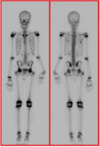
Figure 1: Bone scintigraphy with 99mTc- MDP without evidence of osteoblastic metastatic lesions.

Figure 2: 18F-FDG PET/CT image in the coronal sections showing the bone lesions, one in the left femur (blue arrow) and the other in the proximal epiphyseal plate of the right tibia (red arrow).
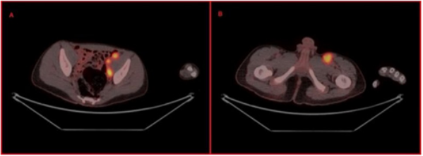
Figure 3: 18F-FDG PET/CT image in the axial sections presenting hypermetabolic lymph node enlargement in the left external iliac (A) and left inguinal (B) chains
Discussion
Retinoblastoma is the most common ocular malignancy and its incidence is 1 in 15,000-20,000 live births [1]. In about 40% of the cases, retinoblastoma is bilateral and all of them are hereditary associated to mutations on gene RB1, localized in chromosome 13q1.4 [1]. Examination of ocular fundus determines the diagnosis and ultrasound, magnetic resonance imaging (MRI) and computed tomography (CT) complement the assessment by differentiating it from other ocular diseases like Coats disease [1,2]. Besides ocular involvement, soft tissue of the orbit, cervical lymph nodes, bone, bone marrow and central nervous system metastases may occur and generally, they are accompanied by high mortality rate mainly at in developing countries where the diagnosis is late [1]. Currently, 18F-FDG PET/CT has been showed an interesting tool to available r e t i n o b l a s t o m a . Radhakrishnan et al showed prognostic value of 18F-FDG PET/CT optic nerve uptake at baseline and response after neoadjuvant chemotherapy in International Retinoblastoma Staging System (IRSS) stage III patient [3]. Moreover, 18F-FDG PET/ CT have higher sensitivity to detect bone metastases did not see at bone scan [3], as observed in our case. Other possibility is to evaluate soft tissue metastases, mainly whole-body MRI has not been available. Based in our case and in its value prognostic [3], new studies using 18F-FDG PET/CT for staging of advanced retinoblastoma could showed its real significance.
References
- Aerts I, Lumbroso Le Rouic L, Gauthier Villars M, Brisse H, Doz F, et al. (2006) Retinoblastoma. Orphanet journal of rare diseases 1(1): 31.
- De Graaf P, Göricke S, Rodjan F, Galluzzi P, Maeder P, et al. (2012) Guidelines for imaging retinoblastoma: imaging principles and MRI standardization. Pediatric radiology 42(1): 2-14.
- Radhakrishnan V, Kumar R, Malhotra A, Bakhshi S (2012) Role of PET/ CT in staging and evaluation of treatment response after 3 cycles of chemotherapy in locally advanced retinoblastoma: a prospective study. Journal of Nuclear Medicine 53(2): 191-198.
