 Case Report
Abstract
The white cerebellum sign is an ominous but infrequent radiological imaging finding encountered commonly in patients with severe often irreversible anoxic-ischaemic brain damage. Recognition of this sign by the radiologists is imperative as it is often associated with an unfavourable prognosis. We report the case of an 8-month old child who presented with severe head injury and brain computed tomography scan done on her revealed the white cerebellum sign. We discussed the relevance of this sign as it affects diagnosis, treatment and prognosis and equally reviewed the pertinent literatures.
Keywords: Anoxic Ischaemic Brain Damage; Prognosis; White Cerebellum Sign
Introduction
Diffuse hyperdensity of the infratentorial brain including thalamus, bainstem and cerebellum with relative hypodensity of the supratentorial brain seen on computed tomography images in severe head injury patients is the “white cerebellum sign.”1 It is also known as “reversal sign” or “dense cerebellum sign” and is often an indicator of severe irreversible anoxic-ischaemic brain damage with a poor prognosis [1-4]. However, the “reversal sign” proper denotes reversal of the gray matter and white matter densities of the cerebral hemisphere with associated increase in the density of thalamus, brain stem and cerebellum [1] Both signs are harbingers of severe irremediable hypoxic brain insult. The general loss of density of the supratentorial brain is ascribed to widespread edema with resultant loss of the grey-white matter interface [5]
The white cerebellum sign is mainly documented in pediatric patients with hypoxic brain damage. About 10-20 % of adult patients with severe head injuries develop generalized brain swelling and in children it is about 20-40 % as common when compared to adult patients [6] White cerebellum sign can be found in patients with the following conditions:- severe head injury, birth sphyxia, drowning, smoke inhalation, status epilepticus, bacterial meningitis, encephalitis [7] and post cardiac arrest hypoxia [1]. It can also be seen in patient with non-accidental hypoxic brain damage [1]. The white cerebellum sign has drawn the attention of scientists over the years but scarcely documented in the literature [8]. There is dearth of information on this sign in our setting as shown by literature search thereby making this article indispensable for both the radiologists and clinicians. Radiologists especially must take cognizance of the classic appearance of the white cerebellum sign so as to make accurate diagnosis and guide management properly as prognosis is almost always hopeless.
Case Report
The patient is an 8-month old female referred to a private diagnostic centre for brain computed tomography on account of severe head injury which parents claim was as a result of fall from a table, a height of about 90cm(3ft). The scan showed diffuse decrease in density of the supratentorial brain with relative increased attenuation of the thalami, brainstem, and cerebellum giving the “white cerebellum sign”. There is also obliteration of the surface and central CSF spaces as well as loss of the gray white matter interface in keeping with severe oedema. No fracture was seen (Figures 1-3).
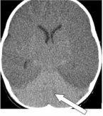
Figure 1: Axial CT image showing the white cerebellum sign (Arrow).
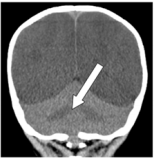
Figure 2: Coronal CT image showing the white cerebellum sign (Arrow).
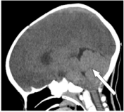
Figure 3: Sagittal CT image showing the white cerebellum sign (Arrow).
Discussion
Although the white cerebellum sign is important from diagnostic, therapeutic and prognostic viewpoint, yet it is an uncommonly described sign in diagnostic radiology [9] The sign portends an irreversible brain damage and was initially described in cases of child abuse, though not definite it can be the only evidence of nonaccidental head injury [9,10] It has also been seen in other cases of extra-cerebral hypoxia like in smothering or strangulation [10] Han et al. [1], in their work analyzed 20 cases of white cerebellum sign in children and reported that trauma is a rare cause of this sign. It has been documented that about one-third of patient with this sign on CT scan will die while the remaining population will suffer irreversible brain damage with subsequent development of diffuse atrophy and cystic encephalomalacia [5].
The hallmark of the white cerebellum sign is intracranial hypertension [4,7,11,12] However the exact mechanism and pathogenesis remain unclear, though several theories have been proposed [1,4,5] Some of which include;
a) Raised intracranial pressure causes partial venous obstruction resulting in distension of deep medullary veins [3].
b) Preferential flow to posterior circulation [2].
c) Transtentorial herniation partially relieving the increased intracranial pressure, and thus increase perfusion of central structures [1,3].
d) Hypoxia limits ATP production and disrupts the sodium pump. The influx of water and sodium into cells causes cytotoxic oedema. Later, the water content rises in the extracellular and intracellular spaces (vasogenic oedema). Low density changes appear in the areas most vulnerable to hypoperfusion [3].
e) Anoxia and ischaemia elevate brain glucose. This hyperglycaemic state preferentially damages the cortex and basal ganglia [1,3].
The treatment of patients with white cerebellum sign is basically symptomatic aimed at reducing intracranial hypertension and reversing ischaemic injury [13, 14] The white cerebellum sign is not uniformly hopeless as earlier reported [15]. Some patients who were found with this sign have been treated successfully and they made significant improvement especially those that followed Meningitis [8,15] At least one patient has been reported to have made full recovery following treatment [15].
Conclusion
The white cerebellum sign is a subtle but classic radiological imaging finding denoting irreversible brain insult with almost dismal outcome. Its recognition is important for the radiologists and clinicians in the managements, monitoring and counseling of these patients.
Acknowledgement
We sincerely thank Dr Okpaku NF, a Consultant radiologist for his assistance throughout the preparation of this manuscript.
References
- Han BK, Towbin RB, De Courten Myers G, McLaurin RL, Ball WS Jr (1989) Reversal sign on CT scan: Effect of anoxic/ischemic cerebral injury in children. AJNR Am J Neuroradiol 10(6): 1191-1198.
- Chavhan GB, Shroff MM (2009) Twenty classic signs in neuroradiology: A pictorial essay. Indian J Radiol Imaging 19(2): 135-145.
- Vergote G, Vandeperre H, De Man R (1992) The reversal sign. Neuroradiology 34(3): 215-216.
- Bird CR, Drayer BP, Gilles FH (1989) Pathophysiology of ‘reverse’ edema in global cerebral ischemia. Am J Neuroradiol 10(1): 95-98.
- Dwarakanath S, Bansal A, Rudrappa S, Gopal S, Venkatramana NK (2006) White cerebellum sign - A case report and review of literature. J Pediatr Neurosci 1(3): 22-23.
- Osbom AG (1994) “Craniocerebral Trauma”; In: Osbom AG (Eds.), Diagnostic Neauroradiology, 1st Edition, Chapter. Elsiver pp. 199-247.
- Shroff Manohar M, Govind B Chavhan (2009) Twenty Classic Signs in Neuroradiology: A Pictorial Essay. Indian Journal of Radiology and Imaging 19(2): 135-145.
- Serge Eddy Mba, Kazadi Kalangu, Aaron Musara, Dumisani Ndlovu, Nathaniel Harunangoni Zimani (2018) White Cerebellum Sign After Ventriculo-Peritoneal Shunt Insertion: A Case Report and Review of the Literature. International Journal of Neurosurgery 2(2): 31-34.
- Malik V, Murthy TV, Raj V, Vyas S, Mehar US (2015) White Cerebellar Sign in Immediate Post-Partum Period. Med J Armed Forces India 71(1): S163-S165.
- Harwood Nash DC (1992) Abuse to Pediatric Central Nervous system. AJNR Am J Neuroradiol 13(2): 569-575.
- Huisman TA, SH Kubat, BP Eckhardt (2007) The Dark Cerebellar Sign. Neuropediatrics 38(3): 160-163.
- Bhoil, Sabina, Rohit Bhoil (2015) Reversal Sign: A Red-Flag in Emergency Departments. Emergency Nurse 23(7): 24-25.
- Divya Singh, Amit Shama (2018) White Cerebellum Sign: Red Flag in the Emergency 16(5): 131213.
- Kjos BO, Brant Zawadzki M, Young RG (1983) Early CT Findings of Global Central Nervous System Hypoperfusion. AJR Am J Roentgennol 141(6): 1227-1232.
- Chalela JA, Rothlisberger J, West B, Hays A (2013) The white cerebellum sign: an under recognized sign of increased intracranial pressure. Neurocrit Care 18(3): 398-399.
