info@biomedres.us
+1 (502) 904-2126
One Westbrook Corporate Center, Suite 300, Westchester, IL 60154, USA
Site Map

Received: October 16, 2023; Published: November 10, 2023
*Corresponding author: Hunde Wayuma, Menesibu District Agricultural Office, Mendi Town, West Wallaga, Oromia, Ethiopia
DOI: 10.26717/BJSTR.2023.53.008443
A cross sectional study was conducted from August 2021 to October 2021 in and Around Menesibu district, West Wollega, Oromia, Ethiopia with the objectives of estimating the prevalence of calves coccidiosis and determine the associated risk factors. Accordingly, during the study period, a total of 304 calves fecal samples were purposively collected and examined for the presence of the oocyts of Eimeria by floatation technique using saline solution. The study revealed that an overall of 24.0% (73/304) calves were infested by coccidia during the study period. The risk factors considered were age, sex, body condition and parity of dam. Coccidiosis were predominant (P<0.05) in poor body condition calves and in soft faecal calves. In conclusion, the present finding has demonstrated that coccidia are one of the important pathogens in calves in the study area. Therefore, appropriate monitoring and control of the disease is advisable in the study farms.
Keywords: Calves; Coccidiosis; Menesibu District; Prevalence; Risk Factors
Ethiopia has the largest livestock population in Africa. The total ruminants population for the country is estimated to be about 59.5 million cattle, 30.7 million sheep and 30.2 million goats [1]. The contribution of livestock to the national economy particularly with regard to foreign currency earnings is through exploration of live animal, meat and skin and hides [2]. However, this extensive livestock resource is not exactly exploited because of many constraints, of which poor animal production and management, improper evaluation of public health importance due to various individual parasitic diseases and inadequate knowledge of epidemiology of parasites which otherwise is of great relevance where the distribution of the disease determine the type and scope of control measures to be applied [3]. Gastrointestinal parasite infections are a problem for both small and large scale farms; however, their impact is greater in sub-Saharan Africa. The prevalence of gastrointestinal parasites and the severity of infection vary considerably depending on the genera of helminth and protozoan parasites involved, animal species, local environmental conditions such as humidity, temperature, rainfall, vegetation and management practices [4]. Coccidiosis is a parasitic disease of the intestinal tract caused by microscopic organisms called coccidia and is one of the most common and important disease of cattle worldwide and more than 13 species of Eimeria and one species of Isospora have been described to infect cattle. Of the 13 species recorded, two of the principal pathogens are E. zurnii and E. bovis.
The most common clinical mani-festations include in appetence, weakness, and loss of weight, diarrhea, depression and anemia [5]. Coccidia parasites are generally host-specific parasites, and very specific to a particular region in the intestines [6]. Coccidiosis in cattle is particularly a problem of confined animals kept under intensive husbandry practices. The disease is more common in housed animals than in those on pastures [5]. Previous works which were conducted in different parts of Ethiopia showed that coccidiosis is a paramount important protozoan disease in younger calves which were kept in poor hygienic status as well as improperly nourished with colostrum [7-9]. Although, coccidiosis is an important cause of calf morbidity and mortality in Ethiopia in general, and in the study area in particular, there is no previous detail information on its prevalence coccidiosis in the study area. Therefore, this study will be conducted to determine the prevalence and associated risk factors of calf coccidiosis in Haramaya University dairy farm, Ethiopia. Dairy cattle are important contributors to the society by providing milk and generation income. However, the economic importance of gastrointestinal parasites especially coccidiosis occurring in calves causes substantial economic losses by reducing the growth rate, production and productivity, and causing calf mortalities. Even though there was a huge number of dairy calves population in Haramaya University dairy farm, eastern Ethiopia, no studies had been so far carried on estimate prevalence of coccidiosis and the associated risk factors in calves of Haramaya University dairy farm. Therefore the overall objective of the study was estimating prevalence of coccidiosis of calves and thereby to assess the associated risk factors in study area.
Description of Study Area
The study was conducted in and around Mendi town, which is found in western Wollega zone of Oromia regional state, Ethiopia. This area has a latitude and longitude of 9048’N and 35o6’E respectively and an elevation of 1583 meters above sea level. It is the administrative center of ManasibuWoreda. Manasibu is bordered on the south by Jarso, on the South West by Begi on the North by the BenishangulGumuz Region and on the south west by Nedjo. The town is approximately 596 km away from Addis Ababa to west direction [10].
Study Animals
The study animals were calves of different age groups categorized into two age groups as group I = 1 to 6 months age and group II = >6 to 12 months age which was determined by asking the owner of the animal orally. Examined animals were also categorized into three according to their body condition: good, medium and poor. This is based on different body visible bone structure and fat deposit [11].
Determination of Sample Size
The sample size required for this study as calculated based on sample size determination method previously described by [12].
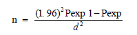
Where, N=required sample size,
Pexp= expected prevalence, d= desires absolute precision.
Accordingly, based on expected prevalence of 72.8% in Mekele town [13], with 5% absolute precision at 95%confidence level, the sample size as determined was 304 calves.
Sampling Techniques and Sample Collection
The sample was collected from calves from Menesibu district. Detailed examination of animals that were selected as samples from selected area was done and convenience to different body condition of animals and different sex and age group (young and adult).
Study Design
A cross sectional study was carried out from August 2021 to October 2021 to determine prevalence of calves coccidiosis by collecting data on events associated with coccidiosis in calves of Menesibu district.
Data Collection
Fecal samples were collected from selected calves after proper restraining of the sampling animal. Fecal samples were collected from rectum by wearing arm length glove through aseptic method. During sampling each sample was properly labeled about relevant information before transporting. Then samples were transported to the Veterinary parasitology laboratory for fecal examinations by simple flotation technique with saline solution [14].
Parasitological Technique
A 5 g portion of each of the collected fecal samples from the total of 30 g was weighed out using a balance and put in a 50 ml beaker. 42 ml of water was added, mixed thoroughly and poured into a 100-ml glass beaker through a strainer. The 50-ml glass beaker was rinsed with 8 ml of water and the total fluid was poured into four 15-ml conical tip centrifuge tubes. After centrifugation at 1,500 rpm for 5 min, the supernatant was decanted and a sodium chloride solution (specific gravity 1.25) was added to the sediment, until the tube was about half full. The content of each test tube was thoroughly mixed with a wooden applicator stick. With the aid of a conical flux, more sugar solution was added until a convex meniscus was form on top of the tube. A glass cover slip was placed on top of each tube and was left for 30 min. Then, each glass cover slip was briskly lifted up and placed on a clean glass slide, not allowing formation of air bubbles. The entire area under each cover slip was examined under a binocular microscope at 400× magnification [14].
Data Analysis
The collected data was first entered and managed into Microsoft Excel worksheet and analyzed by a statistical software namely, SPSS version 22. Prevalence was determined by the formula described by [12] as the rate of number of infested calves and total number of calves in population. Associations between explanatory variables (breed of animals, age, sex, body condition and consistency of feces) and prevalence were done by chi-square test and P<0.05 was set to indicate significance.
Out of all 304 samples, a total of 73 samples (24%) tested positive for Eimeria oocysts. The overall prevalence of coccidiosis in male and female calves was 23.6% and 24.6%, respectively. The overall prevalence of coccidiosis according to age was 25.1% and 32.9% in <6 months and > 6 months of calves respectively. In general, the overall prevalence of coccidiosis were not significantly (P>0.05) according to age and sex of calves. According to body condition score coccidiosis was higher prevalent in poor body condition score than good body condition score calves (P<0.05). Additionally prevalence of Eimeria has revealed that there is a significant association (P<0.05) between the occurrence of Eimeria with parity of dam and fecal consistency (Table 1).
Table 1: Prevalence of Coccidiosis in Calves based on studied risk factors.

Note: Key: n= number of animals examined, *significance difference
The result of the present study showed an overall prevalence of 24.0% in the study area. This finding is comparable with the report of [9] who reported prevalence of 26.04% in calves. This present result was lower than the report of [15] who reported 68.1% prevalence. This variation is most likely attributed to the differences in agro-ecology, and husbandry practices of the study animals in different countries [5]. There was no statistically significant difference in prevalence of coccidian infection between male and female animals (χ2= 0.02, p =0.892), which is in agreement with the reports of [9,15]. This might be associated with the fact that different sex groups kept in similar husbandry system might have equal chance of accessing the oocyts. Despite this, previous studies done on adult cattle showed higher prevalence of Eimeria in female animals than in males [16]. There was statistically significant (χ2=3.95, P=0.047) difference in prevalence rate between fecal consistency and prevalence of coccidiosis which agrees with the findings of [17]. However, this finding disagrees with the report of [15]. Additionally, there was statistically significant difference in prevalence of coccidian infection between parity of dam (χ2 = 10.1, p =0.001), which is disagree with the reports of [17]. A strong significant association of coccidiosis prevalence was recorded between body condition score in the current study. Similarly, [9] reported a higher infection rate in calves with poor body condition score than in calves with good and moderate body condition score. This might be due to the weak immune status of the calves with poor body condition score. As a result, malnutrition and other parasitic infections result in immune compromised calves. This condition produces a higher infection rate in poor state animals than in good-state animals [5]. However, this finding disagrees with the report of [7].
Present study showed that coccidia are infesting significant proportions of calves in the study area. It was shown that poor body condition calves were highly infested with coccidian infection than good body condition calves. All age groups and both sex of calves were found infested with various type of coccidial species. Results from this study indicate the Eimeria infection has a great significance for the livestock producer and need a serious control and preventive issue. Based on the findings of the current study, the following recommendations are suggested:
1. Good feeding and management practices should be put in the farm.
2. Strict awareness on animal husbandry practices should be given to the stakeholders .
3. Further epidemiological investigation on coccidia species should be needed in the study area.


