info@biomedres.us   +1 (502) 904-2126   One Westbrook Corporate Center, Suite 300, Westchester, IL 60154, USA   Site Map
ISSN: 2574 -1241

Impact Factor : 0.548

  Submit Manuscript

Research ArticleOpen Access

Protective Property of Ethanol Extract of C. Sativus on STZ-Induced Diabetic Rat Pancreas Volume 52- Issue 2

Abu OD1*, Ojo I2 and Awhin EP3

  • 1Department of Biochemistry, Faculty of Life Sciences, University of Benin, Nigeria
  • 2Thermo Fisher Scientific Ltd., Carlsbad California, USA
  • 3Department of Medical Biochemistry, Faculty of Basic Medical Sciences, Delta State University, Nigeria

Received: August 08, 2023;   Published: August 18, 2023

*Corresponding author: Abu OD, Department of Biochemistry, Faculty of Life Sciences, University of Benin, Benin City, Nigeria

DOI: 10.26717/BJSTR.2023.52.008233

Abstract PDF

ABSTRACT

The present study investigated the protective property of ethanol extract of Cucumis sativus on streptozotocin (STZ)-induced diabetic rat pancreas. Male albino rats of Wistar strain (n = 25, mean weight = 215 ± 15 g) were assigned to five groups (5 rats/group): normal control, diabetic control, metformin, extract (200 mg/ kg body weight, bwt) and extract (300 mg/kg bwt) groups. A single intraperitoneal injection of 50 mg/kg bwt STZ was used to induce diabetes mellitus in the experimental rats. The diabetic rats were treated for a period of 21 days with 50 mg/kg bwt metformin (standard antidiabetic agent) or the extract. Activities of catalase, superoxide dismutase (SOD), glutathione peroxidase (GPx) and glutathione reductase (GR) as well as glutathione (GSH), total protein (TP), malondialdehyde (MDA) and nitric oxide (NO) levels were measured in pancreatic tissue. The results showed that the diabetogenic agent STZ significantly increased the fasting blood glucose (FBG) concentrations of the rats, but it decreased the activity/concentration of antioxidant enzymes/molecules (p < 0.05). However, treatment of the diabetic Wistar albino rats with 200 and 300 mg/kg bwt extract markedly reduced the FBG concentration and body weights of rats, but enhanced the activity/concentration of antioxidant enzymes/molecules in pancreatic tissue (p < 0.05). These results indicate that ethanol extract of C. sativus fruit has the potential to promote antioxidant defense in STZ-induced diabetic rats pancreas.

Keywords: Cucumis Sativus; Glutathione; Lipid Peroxidation; Medicinal Plant; Oxidative Stress

Abbreviations: SOD: Superoxide Dismutase; GPx: Glutathione Peroxidase; GR: Glutathione Reductase; GSH: Glutathione; TP: Total Protein; MDA: Malondialdehyde; NO: Nitric Oxide; FBG: Fasting Blood Glucose; ROS: Reactive Oxygen Species; AP: Acute Pancreatitis; O-GlcNAcase: O-linked β -N-Acetylglucosaminase

Introduction

Pancreas, a glandular organ in the digestive and endocrine systems of vertebrates, is located in the abdominal cavity behind the stomach. As an endocrine gland it synthesizes insulin, glucagon, somatostatin, and pancreatic polypeptide [1]. The pancreas is also a digestive organ, secreting pancreatic juice which contains enzymes that facilitate digestion and absorption of nutrients in the ileum. Anatomically, the pancreas is divided into a head, which rests within the concavity of the duodenum, a body lying behind the base of the stomach, and a tail, which ends abutting the spleen. Lying anterior to the superior mesenteric artery and vein, the neck of the pancreas resides between the body and head. The head of the pancreas surrounds these two vessels, and a small uncinate process emerges from its lower part, lying behind the superior mesenteric artery [1]. The pancreas has an internal hormonal role (endocrine) and an external digestive role (exocrine). It has two major ducts, the main pancreatic duct, and the accessory pancreatic duct [2]. The functional state of the pancreas is evaluated via determination of the activities of α-amylase, lipase and elastase (in stool) [2]. Certain drugs have been demonstrated to cause acute pancreatitis [3,4]. The role of oxidative stress in disease pathogenesis is well documented. Oxidative stress-triggered mitochondrion membrane rupture leads to pancreatic cell necrosis.

Reactive oxygen species (ROS) produced within the cells rupture lysosomal membrane releasing hydrolases [3,5]. Pancreatotoxicity is characterized by widespread alterations of membranous organelles of acinar cells, especially the endoplasmic reticulum and zymogen granules. Hemorrhagic necrosis of the pancreas is due to endogenous, intraparenchymal activation of zymogen proteases, including proelastase [5]. The necrotic event may also be characterized by severe congestion and edema of the stroma, which contains large quantities of extravasated red and inflammatory cells (mostly neutrophils).

Diabetes mellitus is generally induced in laboratory animals by several methods: chemical, surgical and genetic (immunological) manipulations. Chemical method include the use of STZ, alloxan and sucrose load. Synthesized by a strain of the soil microbe Streptomyces achromogenes (Gram-positive bacterium) STZ is a permanent diabetes inducing drug [4]. Induction of diabetes mellitus with STZ involves damage to the pancreas. This study investigated the protective property of ethanol extract of C. sativus on STZ-induced diabetic rat pancreas.

Materials and Methods

Chemicals and Reagents

All chemicals and reagents used in this study were of analytical grade and they were products of Sigma-Aldrich Ltd. (USA).

Plant Extraction

Freshly harvested C. sativus fruits were purchased from a major vegetable market in Benin City, Nigeria and identified at the Department of Plant Biology and Biotechnology, University of Benin, Benin City, Nigeria. They were washed and air-dried for 4 weeks at the Department of Biochemistry. The dry plant was ground with a mechanical blender. The pulverized sample was cold macerated in absolute ethanol for three days (72 h) in a bell jar and filtered using Whatmann filter paper No. 42 (125 mm). The ethanol extract was thereafter concentrated using rotary evaporator and freeze-dried using a lyophilizer [6-10].

Experimental Animals

Mature male Wistar albino rats (n = 25, mean weight = 215 ± 15 g) were obtained from the Department of Anatomy, University of Benin, Nigeria and housed in wooden cages. They were acclimatized for two weeks before commencement of the study and had free access to feed and clean water.

Experimental Design

The rats were assigned randomly to five groups (5 rats/group): normal control, diabetic control, metformin, extract (200 mg/kg bwt) and extract (300 mg/kg bwt) groups. A single intraperitoneal injection of 50 mg/kg bwt STZ was used to induce diabetes mellitus in the experimental rats. Subsequently, the diabetic rats were treated for a period of 21 days with either metformin (50 mg/kg bwt) or the medicinal plant extract.

Biochemical Analyses

At the end of 21-day treatment, the rats were euthanized under mild chloroform anaesthesia (after an overnight fast). Their pancreases were excised, blotted dry and used to prepare 20 % tissue homogenate. The homogenate was centrifuged at 2000 rpm for 15 min to obtain clear supernatant which was used for biochemical analysis. The activities of catalase, SOD, GPx and α-amylase were determined [11-14]. Levels of pancreatic TP, MDA, GSH, and NO were also measured [15-18]. The activity of GR was determined using a previously described method [19].

Data Analysis

Data are presented as mean ± SEM (n = 5). Statistical analysis was performed using SPSS version 21. Statistical differences between means were compared using Duncan multiple range test. Statistical significance was assumed at p < 0.05.

Results

Effect of Ethanol Extract of C. sativus on Weight and Blood Glucose of Rats

As shown in (Tables 1 & 2), STZ-induced diabetes mellitus significantly increased the blood glucose concentration of the rats (p < 0.05). However, treatment of the diabetic rats with extract of C. sativus fruit markedly reduced their FBG concentration and body weights (p < 0.05).

Table 1: Effect of Ethanol Extract of C. sativus on Weight and Blood Glucose Parameters.

biomedres-openaccess-journal-bjstr

Note: Data are weight and FBG parameters and are expressed as mean ± standard error of mean (SEM, n = 5).

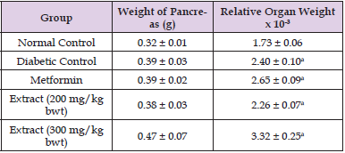
Table 2: Comparison of Organ and Relative Organ Weights.
biomedres-openaccess-journal-bjstr

Note: Data are organ/body weight ratio of rat and are expressed as mean ± SEM (n = 5).

Values with superscript “a” are significantly different from the normal control group.

Oxidative Status in Rats Pancreases

Treatment of STZ-induced diabetic Wistar rats with ethanol extract of C. sativus significantly increased the activities of the antioxidant enzymes as well as concentrations of GSH and NO, but it markedly reduced the activity of α-amylase and concentrations of pancreatic TP and MDA (p < 0.05). These results are shown in (Tables 3-6).

Table 3: Effect of Ethanol Extract of C. sativus on Pancreatic Total Protein and Glutathione Levels.

biomedres-openaccess-journal-bjstr

Note: Data are pancreatic total protein and GSH levels and are expressed as mean ± SEM (n = 5).

Values with superscript “a” are significantly different from the normal control group.

Values with superscript “b” are significantly different from the diabetic control group.

Table 4: Effect of Ethanol Extract of C. sativus on Oxidative Status in Rat Pancreas.

biomedres-openaccess-journal-bjstr

Note: Progressive motility (PM); Linearity (LIN); Straightness (STR); Trail speed (VCL); Progressive Speed (VSL); Speed of travel (VAP); Amplitude of lateral head displacement (ALH); Total Motility (MT); wobble index (WOB); Micromeres per second (μm/s).

Table 5: Effect of Ethanol Extract of C. sativus on Pancreatic Alpha- Amylase and Glutathione Peroxidase Activity.

biomedres-openaccess-journal-bjstr

Note: Data are activities of pancreatic α-amylase and glutathione peroxidase and are expressed as mean ± SEM (n = 5).

Values with superscript “a” are significantly different from the normal control group.

Values with superscript “b” are significantly different from the diabetic control group.

Table 6: Effect of Ethanol Extract of C. sativus on Pancreatic NO Level.

biomedres-openaccess-journal-bjstr

Note: Data are pancreatic NO concentrations and are expressed as mean ± SEM (n = 5).

Discussion

Acute pancreatitis (AP) may be caused by exposure to certain drugs/compounds [3] Literature has shown that about 20 % of the top most prescribed drugs are toxic to the pancreas [4,5]. Some causes of pancreatitis are gallstones, alcohol abuse, hypercalcemia, hypertriglyceridemia, viral infection, trauma as well as cardiovascular and anti-inflammatory analgesic agents [20]. Pancreatotoxicity due to direct exposure to drugs results in subclinical pancreatic damage [21]. Two out of the following three features are required for a diagnosis of AP:

a) Abdominal pain characteristic of acute pancreatitis;

b) Serum amylase and/or lipase levels ≥ 3 times the upper limit of normal; and

c) Results of CT scans [6].

As a localized inflammation of the pancreas the disease is commonly mediated by the premature activation of digestive enzymes retained in the organ. Although this condition may resolve by itself within days, its persistence could cause pancreatic dysfunction and failure of other remote organs/systems [22]. Pancreatitis is of two forms: acute and chronic. It was recently discovered that chronic pancreatitis is a consequence of repeated episodes of an acute case [23]. One of many chemical agents that can damage the pancreas is STZ. Induction of diabetes mellitus with STZ causes serious damage to the organ [11]. The exact molecular mechanism underlying the cytotoxic effect of STZ is not well-understood, however studies suggest that the cytotoxicity could be via production of reactive oxygen species (ROS) thus inducing oxidative stress, causing DNA damage with resultant necrosis (due to the DNA methylating activity of the methyl nitroso urea moiety of the drug), release of NO which inhibits aconitase activity resulting in mitochondrial dysfunction, or via inhibition of O-linked β -N-acetylglucosaminase (O-GlcNAcase) [24]. Increasing evidence suggest that oxidative stress plays a role in the pathogenesis of diabetes mellitus and its complications [25]. Hyperglycemia increases oxidative stress, which contributes to impairment of processes that fail during diabetes (insulin action and secretion). In addition, antioxidant mechanisms are diminished in diabetic patients, which may further promotes oxidative stress [26,27].

This study investigated the protective property of ethanol extract of C. sativus on STZ-induced diabetic rat pancreas. The results showed that induction of diabetes mellitus with STZ significantly increased the FBG concentrations of the rats, but it decreased the activity/concentration of antioxidant enzymes/molecules. However, treatment of the diabetic Wistar albino rats with two different doses of ethanol extract of C. sativus fruit markedly reduced the FBG concentration and body weights of rats, but enhanced the activity/concentration of antioxidant enzymes/molecules in pancreatic tissue. These results are in agreement with those of previous studies [28-33]. The results suggest that oxidative stress plays a crucial role in STZ-induced diabetes mellitus and that increased ROS production suppresses the activity of antioxidant enzymes, while diminishing the levels of antioxidant molecules in pancreatic tissue. The beneficial effect of the medicinal plant may not be unconnected to its bioactive compounds. Studies have shown that plants containing important phytochemicals have many pharmacological/biological effects [34-67].

Conclusion

The results obtained in this study indicate that ethanol extract of the medicinal plant fruit has the potential to promote antioxidant defense in STZ-induced diabetic rat pancreas.

References

  1. Drake RL, Vogl W, Tibbitts AWM (2005) Gray's anatomy for students. Philadelphia: Elsevier/Churchill Livingstone 297(303): 288-290.
  2. Young B (2006) Wheater's functional histology: a text and colour atlas. Churchill Livingstone/Elsevier Pp. 299-301.
  3. Kaurich T (2008) Drug-induced acute pancreatitis. Proc Bayl Univ Med Cent 21(1): 77-81.
  4. Badalov N, Baradarian R, Iswara K, Li J, Steinberg W, et al. (2007) Drug- induced acute pancreatitis: an evidence-based review. Clin Gastroenterol Hepatol 5(6): 648-661.
  5. Eltookhy A, Pearson NL (2006) Drug-induced pancreatitis. Can Pharmacists J 139(6): 58-60.
  6. Abu OD, Imafidon KE, Obayuwana HO, Okuofu ED (2017) Phytochemical, proximate, and metal content analysis of citrullus lanatus (watermelon) seeds. FUDMA Journal of Sciences 2(2): 153-156.
  7. Abu OD, Onoagbe IO (2019) Biochemical effect of aqueous extract of Dialium Guineense stem bark on oxidative status of normal Wistar rats. International Journal of Clinical Biology and Biochemistry 1(2): 18-21.
  8. Abu OD, Imafidon KE, Iribhogbe ME (2015) Biochemical effect of aqueous leaf extract of Icacina trichanta Oliv. on urea, creatinine and kidney oxidative status in CCl4-induced Wistar rats. Nigerian Journal of Life Sciences 5(1): 85-89.
  9. Abu OD, Onoagbe IO, Obahiagbon O (2020a) Alpha amylase and alpha glucosidase inhibitory activities of extracts of Dialium guineense stem bark. International Journal of Clinical Biology and Biochemistry 2(1): 7-10.  
  10. Abu OD, Onoagbe IO, Osemwenoyenmwen O (2020b) Alpha amylase and alpha glucosidase inhibitory activities of isolated total saponins and tannins of Dialium guineense stem bark. Journal of Cellular and Molecular Biology Research 1(2): 1-3.
  11. Cohen G, Dembie CD, Marcus J (1970) Measurement of catalase activity in tissue extracts. Analytic Biochemistry. 34: 30-38.
  12. Misra HR, Fridovich I (1972) The role of superoxide anions in the auto oxidation of epinephrine and a single assay for superoxide dismutase. J Biol Chem 247(10): 3170-3175.
  13. Rotruck JT, Pope AL, Ganther HE, Swanson AB, Hafeman DG, et al. (1973) Selenium biochemical role as a component of glutathione peroxidase. Science 179(4073): 588-590.
  14. Bernfeld P (1951) Enzymes of starch degradation and synthesis. Adsan Enrs rnoL 12: 379-428.
  15. Henry RJ, Sobel C, Beckman S (1957) Determination of serum protein by the Biuret reaction. Anal Chem 92(149): 1-5.
  16. Ellman GL (1959) Tissue sulphydryl groups. Archive of Biochemistry and Biophysics 82(1): 70-77.
  17. Guttridge JMC, Wilkins C (1982) Cancer dependent hydroxyl radical damage to ascorbic acid. Formation of thiobarbituric acid reactive product FEBS Lett 137(2): 327-330.
  18. Marcocci L, Packer L, Droy Lefaix MT, Sekaki A (1994) Antioxidant action of Ginkgo biloba extract EGb 761. Methods in Enzymology 234: 462-475.
  19. Abu OD, Ikponmwosa Eweka O (2022a) Evaluation of the Potential of Total saponins and Tannins of Dialium guineense Stem Bark in the Amelioration of Carbon Tetrachloride-Induced Renal Oxidative Stress. SAU Science Tech Journal 7(1): 42-50.
  20. Mallick S (2004) Metformin induced acute pancreatitis precipitated by renal failure. Postgrad Med J 80(942): 239-240.
  21. Schmidt LE, Dalhoff K (2004) Hyperamylasaemia and acute pancreatitis in paracetamol poisoning. Aliment Pharmacol Ther 20(2): 173-179.
  22. Banks PA, Freeman ML (2006) The Practice Guidelines in Acute Pancreatitis. Am J Gastroenterol 101(10): 2379-2400.
  23. Dragovic G, Milic N, Jevtovic D (2005) Incidence of acute pancreatitis and nucleoside reverse transcriptase inhibitors usage. Int J STD AIDS 16(6): 427-429.
  24. Anabel SGR, Shoshan KS, Jan DH, Alphons PLV, Dirk WB (2010) Anomer equilibrated streptozotocin solution for the induction of experimental diabetes in mice (Musmusculus). Journal of the American Association for Laboratory Animal Science 49(1): 40-44.
  25. Brownlee M (2001) Biochemistry and molecular cell biology of diabetic complications. Nature 414(6865): 813-820.
  26. Sanders RA (2003) Diabetes oxidative stress, and antioxidants: a review. J Biochem Mol Toxicol 17(1): 24-38.
  27. Rains JL, Jain SK (2011) Oxidative stress, insulin signaling, and diabetes. Free Radic Biol Med 50(5): 567-575.
  28. Abu OD, Imafidon KE, Obayuwana O (2020) Ethanol leaf extract of Anacardium occidentale ameliorates alloxan-induced changes on blood glucose level and lipid profile of Wistar rats. IAR Journal of Medical Sciences 1(5): 257-262.
  29. Abu OD, Imafidon KE, Obayuwana O (2020) Effect of aqueous extract of Anacardium occidentale leaves on blood glucose level and lipid profile of diabetic rats. Global Scientific Journal 8(10): 977-987.
  30. Okpiabhele AO, Nwanze EAC, Abu OD (2018) Therapeutic Potential of Virgin Coconut Oil in Ameliorating Diabetes Mellitus and Hepatotoxicity Using Rattus Norvegicus as Case Study. Asian Journal of Biological Sciences 11(3): 138-144.
  31. Abu OD, Alegun O, Ifekwe JC (2023) Renal Oxidative Status in Diabetic Wistar Rats Administered Methanol Fraction of Ethanol Extract of Dialium guineense. Medical and Clinical Case Reports Journal 1(1): 12-15.
  32. Abu OD, Awhin EP, Ifekwe JC (2023) Liver Function Status of Diabetic Wistar Rats Treated with Ethanol Extract of Cucumis sativus Fruit. Biomedical Journal of Scientific and Technical Research 51(2): 42440-42445.
  33. Abu OD, Ojo I, Ezike TV (2023) Methanol Fraction of Ethanol Extract of Dialium guineense Stem Bark Mitigates STZ-Induced Oxidative Stress in Rat Liver. Biomedical Journal of Scientific and Technical Research 51(2): 42594 -42600.
  34. Abu OD, Onoagbe IO, Obahiagbon O (2020) In Vitro Antioxidant Activities of Isolated Total Saponins and Tannins of Dialium Guineense Stem Bark. IAR Journal of Medical Sciences 1(4): 193-199.
  35. Abu OD, Onoagbe IO, Obahiagbon O (2020) Vitamin contents of extracts of Dialium guineense stem bark. Biomed J Sci and Tech Res 30(2): 23263-23267.
  36. Abu OD, Onoagbe IO, Obahiagbon O (2020) Analyses of metal and amino acid compositions of aqueous and ethanol stem bark extracts of Dialium guineense. Journal of Biogeneric Science and Research 6(4): 1-3.
  37. Abu OD, Onoagbe IO, Obahiagbon O (2020) Phenolic contents of extracts of Dialium guineense stem bark. American Journal of Sciences and Engineering Research 3(4): 92-96.
  38. Abu OD, Onoagbe IO (2021) Acute toxicity of aqueous and ethanol extracts of Dialium guineense stem bark. Journal of Bioinnovation 10(2): 427-432.
  39. Abu OD, Adeogun EF, Ebhohon SO (2019) Oral LD50 of total saponins and tannins isolated from Dialium guineense stem bark. European Journal of Experimental Biology 9(2): 11-13.
  40. Abu OD, Onoagbe IO, Ojo I (2021) Determination of effective dose for ethanol extract of Dialium guineense stem bark. Journal of Medical Research and Case Reports 3(2): 1-4.
  41. Abu OD, Ikponmwosa Eweka O (2022) Potential of Extracts of Dialium guineense Stem Bark in the Mitigation of Carbon Tetrachloride-induced Renal Oxidative Stress. BIU Journal of Basic and Applied Sciences 7(1): 62-69.
  42. Abu OD, Ikponmwosa Eweka O (2022) Potential of Total Saponins and Tannins Isolated from the stem bark of Dialium guineense in the Amelioration of Kidney Dysfunction Caused by CCl4. Journal of Basic and Applied Medical Sciences 2(1): 1-6.
  43. Abu OD, Onoagbe IO, Ojo I (2021) Determination of effective dose for ethanol extract of Dialium guineense stem bark. Journal of Medical Research and Case Reports 3(2): 1-4.
  44. Abu OD, Onoagbe IO, Ojo I (2022) Dose response of total saponins isolated from the stem bark of Dialium guineense. Journal of Advances in Plant Biology 1(4): 1-6.
  45. Abu OD, Onoagbe IO, Ojo I (2021) Graded and quantal dose response of total tannins isolated from the stem bark of Dialium guineense. Advanced Research Journal of Medicine and Clinical Science 08(10): 699-703.
  46. Abu OD, Onoagbe IO, Ojo I (2022) Dose response study of aqueous extract of Dialium guineense stem bark. American Journal of Biomedical Science and Research 15(2): 252-254.
  47. Abu OD, Imafidon KE, Obayuwana HO (2021) Nephrotoxic and in vivo antioxidant effects of citrullus lanatus seed extract. Biomedical Journal of Science and Technical Research 33(5): 26281-26286.  
  48. Abu OD, Imafidon KE, Obayuwana HO, Onodje S (2020) Quantitative phytochemical evaluation and phenolic contents of extracts of Citrullus lanatus seed. Int J Bioorg Chem Mol Biol 7(1): 31-35.
  49. Abu OD, Ngedaa OS, Osarhenomase EG (2022) Effect of Extracts of Dialium guineense Stem Bark on Lipid peroxidation Index and Histological Changes in Kidneys of Normal Rats. Forensic Medicine 4(2): 23-29.
  50. Abu OD, Onoagbe IO, Ekugum E (2022) Nephrotoxic Evaluation of Aqueous Stem Bark Extract of Dialium guineense in Normal Wistar Rats. Journal of Pharmaceutical and Bio Medical Sciences 2(9): 353-357.
  51. Abu OD, Onoagbe IO, Ohikhuare F (2022) Nephrotoxic Evaluation of Ethanol Stem Bark Extract of Dialium guineense in Normal Wistar Rats. International Journal of Forensic Medicine 4(2): 19-22.
  52. Abu OD, Umar AB, Eiremiokhae CO (2022) Investigation of the Cardioprotective Capacity of Aqueous Extract of Icacina trichanta Leaves in Rats Exposed to CCl4. Journal of Genetics and Cell Biology 6(1): 322-328.
  53. Abu OD, Onoagbe IO, Ekugum E (2022) Hepatotoxicity of Graded Doses of Ethanol Extract of Dialium guineense Stem Bark in Wistar Rats. Journal of Pharmaceutical and Bio Medical Sciences 2(9): 347-352.
  54. Abu OD, Ezike TV, Ajuwa OI (2022) Cardioprotective property of extracts of Dialium guineense stem bark in rats exposed to CCl4. American Journal of Biomedical Science and Research 16(6): 689-693.
  55. Abu OD, Umar AB, Ajuwa OI (2022) Protective Property of Total Saponins and Tannins of Dialium guineense Stem Bark in CCl4-Induced Cardiotoxicity in Rats. World Journal of Genetics and Molecular Biology 1(1): 1-6.
  56. Abu OD, Orobator ON, Momodu IB (2022) Investigation of the Hepatoprotective Effect of Extracts of Dialium guineense Stem Bark in Wistar Rats Exposed to CCl4. Journal of Clinical Gastroenterology and Hepatology 4(2):123-126.
  57. Abu OD, Orobator ON, Momodu IB (2022) Evaluation of the Effect of Total Saponins and Tannins Isolated from Dialium guineense Stem Bark on CCl4 - Induced Hepatotoxicity in Wistar Rats. Global Journal of Medical and Clinical Case Reports 9(3): 035-038.
  58. Abu OD, Eromosele AI, Osarhenomase EG (2022) Effect of Extracts of Dialium guineense Stem Bark on Lipid Profile and CCl4- Induced Histological Changes in Liver of Wistar Rats. International Journal of Lipids 1(1): 22-28.
  59. Abu OD, Omage JI, Ogbebor EO (2022) Effect of Total Saponins and Tannins Isolated from the Stem Bark of Dialium guineense on Lipid Profile and CCl4- Induced Histological Changes in Liver of Wistar Rats. Journal of Medicine and Biology 3(2): 1-9.  
  60. Abu OD, Iyare HE, Ogboi KU (2022) Cardiac Oxidative Status in CCl4-Exposed Rats Treated with Extracts of Dialium guineense Stem Bark. Global Journal of Scientific Frontier Research 22(01): 1-6.
  61. Abu OD, Okuo AV, Osemwota OF (2022) Extracts of Dialium guineense Stem Bark Ameliorates CCl4-induced Oxidative Stress in Liver of Wistar Rats. Biomedical Journal of Scientific and Technical Research 46(2): 37297-37301.
  62. Abu OD, Okuo AV, Osemwota OF (2022) Total Saponins and Tannins of Dialium guineense Stem Bark Protect Against CCl4-induced Oxidative Stress in Rats Liver. International Journal of Medical and Clinical Case Reports 1(1): 15-20.
  63. Abu OD, Ogbebor EO, Omage JI (2022) Effect of Extracts of Dialium guineense Stem Bark on Oxidative Status in Rats Exposed to CCl4. Journal of Clinical Gastroenterology and Hepatology 4(3):124-127.
  64. Abu OD, Iyare HE, Ogboi KU (2022) Antioxidant Property of Total Saponins and Tannins of Dialium guineense Stem Bark in Rats Hearts Exposed to CCl4. Journal of Clinical Epidemiology and Toxicology 3(3): 1-4.
  65. Abu OD, Omage JI, Ogbebor EO (2022) Effect of Total Saponins and Tannins Isolated from Dialium guineense Stem Bark on Oxidative Status in Rats Exposed to CCl4. J Urology and Nephrology 4(4): 500-504.
  66. Abu OD, Avenbuan SE, Ayele P (2022) Investigation of the haematotoxicity of ethanol stem bark extract of Dialium guineense in rats. Journal of Clinical and Biomedical Advance 2(1): 1-4.
  67. Petrov MS, Yadav D (2019) Global epidemiology and holistic prevention of pancreatitis. Nat Rev Gastroenterol Hepatol 16(3): 175-184.