Impact Factor : 0.548
- NLM ID: 101723284
- OCoLC: 999826537
- LCCN: 2017202541
Azize Demirpolat*
Received: October 10, 2022; Published: October 19, 2022
*Corresponding author: Bingöl University/ Food, Agriculture, Animal, Vocational School, Plant and Animal Production Department, Bingol, Turkey,ademirpolat@bingol.edu.tr
DOI: 10.26717/BJSTR.2022.46.007394
The genus Rindera Pall. belongs to Boraginaceae family, tribe Cynoglosseae DC. and includes about 25 species mostly distributed in central and western Asia and in regions with southern and continental climate. In this study, the root and stem anatomical features of the locally endemic Rindera caespitosa (A. DC.) Bunge species distributed in Turkey were investigated for the first time. In anatomical studies, cross-sections were taken for root and stem measurements, stained and examined. The excess of periderm tissue is noticeable in the root cross-section. There are 7-13 layers of cortex under the periderm layer. The parenchyma cells forming the cortex parenchyma show a regular arrangement and consist of flattened cells in the cross section of the stem, cortex parenchyma cells are generally oval and show an irregular arrangement and 8-19 layers. With this study, it is aimed to provide data for systematic studies.
Keywords: Rindera; Root; Stem; Anatomy
The genus Rindera Pall. belongs to Boraginaceae tribe Cynoglosseae DC. and includes about 25 species mostly distributed in central and western Asia. The family Boraginaceae contains about 85 genera and 1600-1700 species of cosmopolitan distribution, except for the wet tropics [1]. Rindera is morphologically characterized by tubular corollas, long anthers with staminal filaments usually inserted at the throat, a style often exserted from the corolla, and large, mostly eglochidiate mericarpids with a broad, membranous wing. All species are perennial and linked to the dry and continental climate of the steppe and semidesertic belts [2]. Rindera is represented by 4 species in Turkey, with the addition of R. cetineri Yıldırım [3], the number of Rindera taxa in Turkey is now increased to 6 species, 3 of are (R. cetineri, R. dumanii Aytaç & R.R.Mill and R. caespitosa) endemic to the Anatolian Peninsula [4,5]. Yıldırım is published Rindera cetineri as a new species, which is restricted to the Akdağ Mountain in the Çameli district of the province of Denizli in western Anatolia, Turkey. It shows morphological similarities to Rindera caespitosa. R. cetineri is easily distinguished from R. caespitosa, especially by its linear to linear lanceolate and light pinkish purple with darker striation lobes; corolla lobes divided into 2/5–1/2 corolla lenght [3]. Rindera caespitosa mostly distributed in provinces of Hakkari, Bitlis, Adıyaman, Bingöl, Elazığ, Erzincan, Erzurum, Kahramanmaraş, Muş, Sivas, Van cities of Turkey [6]. Endemic Rindera caespitosa were silvery grey tomentosa perennial herbs.
The stems simple 5-25 cm, densely leafy. Basal leaves linear-lanceolate, lamina 75-120 x 10-45 mm; cauline linear, very acute, sessile, adpressed tos tem. Inflorescence terminal, corymbose. Calyx lobes 5.5-6.5 mm, lancelotae-ovate, obtuse, 1-nerved. Corolla reddish-purple, 8-12 mm tube equllaing. Stem leaves distinct and lanceolate. Anthers included equal or scarcely protruding corolla lobes. Nutlets a winged and 12-16 mm, suborbicular; wing 3-4 mm broad entire or slightly dentate, wihout hairy [6]. When the studies on Rindera are examined, it is noticed that there are few anatomical studies. Attar et al., 8 genera (Paracarium (DC.) Boiss., Mattiastrum (Boiss.) Brand., Microparacaryum Hilger & Podlech Popov ex H. Riedl., Rindera Pall., Cynoglossum L., Solenanthus Ledeb., Trachelanthus) in the Cynoglosseae (Boraginaceae) tribus Kunze and Lindelofia Lehm.) examined the leaf and stem surface anatomy of 25 species in detail [7]. Karaçoban et al. performed anatomical, micromorphological and karyological studies on the Rindera cetineri species for the first time [8]. In the present paper we provide a brief survey of anatomical features of the Rindera and discuss some aspects that may contribute to a better insight in the difficult taxonomy in Rindera genus and will contribute for further investigation in the future.
Plant Material
Rindera caespitosa was collected from Türkiye, steppe, stony areas, on 3.06.2019, at an altidude of 1500-1550 m. The taxonomic description of the plant was prepared according to Flora of Turkey, volume 6 [6]. Fresh specimens of this new species, defined according to the flora of Turkey, were stored in 70% alcohol for anatomy studies.
Anatomical Study
For the anatomical study, transverse sections from the stem and leaf of the samples prepared with 70% alcohol and superficial sections from the upper and lower surfaces of the leaf were made by hand. Safranin-fast green [9] was used for painting anatomical sections (Table 1, Figures 1-3). Anatomical photography and measurement of the specimens were performed with the help of a digital camera with a Euromex CMEX-10PRO trinocular microscope.
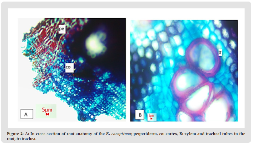
Figure 2 A: In cross-section of root anatomy of the R. caespitosa; pe:periderm, co: cortex, B: xylem and tracheal tubes in the root, tr: trachea.

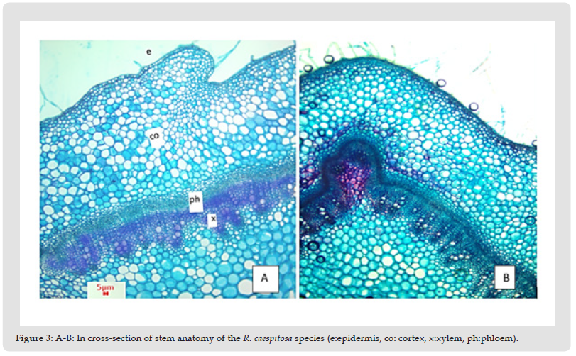
Figure 3 A-B: In cross-section of stem anatomy of the R. caespitosa species (e:epidermis, co: cortex, x:xylem, ph:phloem).

Root
The outermost part of the root is the 3-14 layer of periderm. The periderm is stained red in the root section. There are 7-13 layers of cortex under the periderm layer. The parenchyma cells forming the cortex parenchyma show a regular arrangement and consist of flattened cells. The phloem layer occupies a narrow area and the cell sizes are smaller. The cambium is in the form of a continuous layer with two rows of cells. There are xylem elements under the phloem. The xylem consists of trachea and tracheid elements and is distributed in the pith region. The pith region is lost and covered with xylem (Figure 2).
Stem
The stem is composed of single layered and consists of mostly ovoidal and some cells square-like. In addition, the body is covered with dense hairs originating from the epidermis layer. The hairs are very dense not only on the stem, but also on the leaves. Under the epidermis, there are 1-3 collenchyma layers with different cell shapes. The cortex layer consists of collenchyma cells and cortex parenchyma cells. Cortex parenchyma cells are generally oval and show an irregular arrangement and 8-19 layers. There are phloem cells under the cortex parenchyma. The phloem layer covers a narrower area than the xylem. Between the phloem and the xylem is the cambium. Phloem is composed of 7-12 layers of cells. Xylem is composed of 10-23 layers of cells. The xylem consists of trachea and tracheid elements and the cell walls show thickening. There is no pith in the cross-section of the stem of the plant. The inner part of the stem of the plant is empty (Figure 3).
Attar, et al. [7] examined the stem and leaf anatomy of R. lanata, R. albida and R. bungei Gürke species in their study. R. lanata and R. albida species are characterized by having isobilateral leaves. They observed epidermis, palisade and sponge parenchyma in leaves. In the stem, they observed collenchyma consisting of parenchymatic cells and 4-5 layered cortex under the epidermis, and phloem and xylem under the cortex. The pith consists of large and cylindrical parenchymatic cells [8]. In our study, there is no pith in the cross-section of the stem of the plant. The inner part of the stem of the plant is empty. According to the data obtained from another study, when the root anatomy of the R. cetineri species is examined, there is a fragmented periderm layer in the outermost layer. Under the periderm layer is the cortex layer. The cortex layer consists of 10-12 rows of parenchyma cells. There are phloem cells under the parenchyma cells. The xylem is scattered in the pith region and the pith region has disappeared [8]. In our study, the features of the cortex layer in the cross section taken from the root and the fact that the core area is covered with xylem were found to be similar to this study. However, the periderm layer was quite thick in our study. In the taxonomic evaluation of plants, morphological, anatomical studies of the species are of great importance. The stem and root anatomy of the R. caespitosa species was determined through this study. Our study provides taxonomic data for future anatomical studies [9].


