 Research Article
Abstract
Along a series of previous contributions for Anisotropic Analytic Model (AAA) improvements, several exact/approximated formulations/corrections for wedge filters (WF) photon-dose delivery were presented. Namely, exact photon-beam pathlength through wedge, exact beam limit divergence angle algorithm, and dose delivery correction Omega Factor for wedge filters. Based on all these algorithms/software, a number of 3D comparative-optional simulations with several software systems are developed for AAA model 18 Mev photon-beam. Explicitly, Matlab, Freemat and GNU Octave systems. In these new simulations algorithm, the corrected AAA photon beam intensity I(z) magnitude modification, fluence and geometrical Omega Factor are implemented. Results comprise a series of optimized 3D graphics with numerical data for AAA photon-dose delivery modified by WF. Computational findings show 3D charts for all these programming systems. New results with 4D Interior Optimization imagingdevelopment are presented with software. Resulting consequences for comparative evaluation of the programming methods with all systems are explained. Radiotherapy Medical Physics applications for wedge filter photon-dose calculations emerge from all the numerical and graphical outcomes. Extrapolated clinical radiotherapy applications are obtained from 2D/3D graphical simulation series.
Keywords: Radiation Dose; Attenuation Exponential Factor (AEF); Simulations; Nonlinear Optimization, Matrix Algebra; Spherical-Spatial Analytical Geometry; Series Approximations; Multi-Leaf Collimator (MLC); Wedge Filter (WF); Conformal Wedge Filter; Anisotropic Analytic Model AAA; Intensity Modulated Radiotherapy (IMRT); Intense Modulated Protontherapy (IMPT); Fluence Factor (FF); Treatment Planning Optimization (TPO)
Introduction
Radiation Therapy has been during recent decades among the most important/frequent clinical methods/treatments for cancer cure, remission, palliative care, lifetime elongation, methastasis treatment, and optimization of oncological therapy for patient lifequality. However, the current tumor treatment trends have evolved/ changed in recent years. Chemotherapy, Inmunotherapy, and new Nanoinmunotherapy have emerged as the most powerful methods to eliminate the tumor, obtain a longer patient lifetime, and in many cases, get complete cure/retardation-usually combined one another with/without radiotherapy. In parallel, and spite of these frontline medical advances, radiation therapy has experienced also excellent innovations for oncological treatments. Among them, IMRT, IMPT, Carbon-Ion Therapy with their variants have got outstanding advances for tumor growth-control/cure, terminal patients, methastasis treatment, and side-effects reduction [1-8].
Grosso modo, the progresses in cancer therapy can be classified into two groups—which are synergic, complementary and interactive each other. First one is the science-strictly physics, chemistry, bio/molecular-chemistry, pharmacology, and biology group. Second is the computational, imaging, and software framework that is integrated in oncology to optimize, speed up and make efficient all the advances of the first one— for instance, the incorporation of the Artificial Intelligence for radiotherapy treatment planning optimization/selection, TPO. All these improvements are reduced for the increasing difficulty to control the rising incidence/prevalence of almost all tumor types due to multiple factors apart from lifetime elongation. As a prehypothesis that has been explained in previous contributions [1- 7], Radiation Therapy will remain in clinical future to complete the primary attack to eliminate the most tumor volume and open the field for subsequent Chemo-Inmuno Therapy stages. Complementary, Preventive Medicine has a significant role for cancer incidence reduction and early-stage diagnosis. In a series of previous contributions [1-7], Radiation classical photon therapy AAA model developed by Ulmer and Harder, [1-11], was improved/ complemented.
The studies group along 2008-present [1-7], proved a series of geometrical improvements for WF dose delivery with AAA model. These are geometrical Omega Factor, exact path through WF and limit angle for photon-beam through WF [1-7,9-21]. These mathematical formulation developments are based on analytic geometry, integral equations calculus and programming methods [1-8]. Superposition-convolution photon models constitute the base for analytical proton beamlet model for proton therapy dosimetry [12]. This article is focused on 3D computational simulations for WF 18 Mev beam photon-dosimetry with AAA model. The aim is to demonstrate the efficacy/utility of the presented simulations for treatment planning optimization. Software method was done with three different programming systems. Namely, Matlab, GNUOctave and Freemat. Results comprise a series of 3D imaging and numerical simulations with beam intensity factor I(z) and beamfluence corrections.
Additionally, a comparative assessment of these computational systems for 3D image processing is shown. Consequently, the novelty of this study comprises several strands. The most important is to prove the utility of the 3D planning optimization/ simulation with imaging processing methods. The second is to demonstrate that this utility is efficacious and can be achieved with a number of computational systems available today. Third is the implementation in 3D model simulations of I(z) factor corrections [9]. Complementary, the research shows the usage of these methods for investigation, planning system improvements, and hospital oncology services medical-treatment advances. Therefore, the contribution to medical physics literature can be obtained from these presented results. In summary, this study reports a software programming series of methods to carry out 3D radiotherapy simulations for dose delivery with AAA model and 18 Mev photon spectrum. A number of approximations were applied, but the main objective to prove the utility of 3D dose graphical simulation was reached. The second aim is to show how this can be performed with different computational systems. Namely, Matlab, GNUOctave and Freemat. Medical Physics applications both for clinical radiotherapy treatment planning optimization and radiotherapy physics modelling research come from all the article results.
Mathematical and Computational Methods
Section is divided into two sub-sections. First the AAA model mathematical framework is presented. Secondly, the algorithms computational software and program implementation is explained.
Mathematical and Algorithmic Development
Along a series of papers, [1-11], the equations of the AAA original model photon-dose with WF were calculated. AAA model was named Superposition-Convolution for its mathematical formulation. That is, the term ‘Superposition’ comes from the sum of three Gaussians into the integral. The term ‘Convolution’ describes the mathematical transformation carried out into the Dose-Deposition Kernel at the Integral. Based on this number of previous articles, [1-7,16-29], the WF dose delivery in water without tissue-attenuation corrections is,
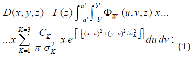
where I(z) is the area integral of the dose over a plane perpendicular to the z-axis at depth z, normalized to one incident electron, σ(z) is the depth-dependent mean square radial displacement, x, y, z are the coordinates of the dose-delivery point at beam-output coordinates system, u, v, z, are the coordinates of photon-fluence at depth z, a, b, are the field-size magnitudes at depth z, CK are optimization parameters resulting for a triple Gaussian function setting, ΦW is the photon fluence modified for WF, a’(z) =a(l + z/F), b’(z) =b(1 + z/F) are the halfside lengths projected into depth z, with F as the source-surface distance (SSD). All these parameters are defined in [30,31].
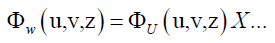
The modified fluence factor (FF) for WF reads,
 with Φw (u,v,z)as WF modified
fluence. (2)
with Φw (u,v,z)as WF modified
fluence. (2)
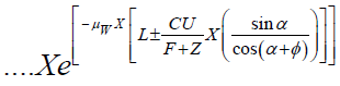
where C is the distance in z-coordinate from source to WF surface, L is half-length of WF for y coordinate, and ΦU = Φ0 / (1 + z/F)2 according to [30,31]. The source fluence is modified primarily for the dose-delivery depth z and secondly by the WF parameters [1- 11, 20,21]. That is, WF modifies the photon-beam energy spectrum. This model of WF was proven be not totally exact [1-7], because the exact path through WF was calculated approximately [1-7]. Then, in [1-11,20,21], the demonstration of the approximate path and exact path was presented. It was determined an Omega correction Factor (OF) for the Equation 2 [1-7] as follows,
• Proposition 1 [Casesnoves, 2014, 1-7]. - Geometrical Omega Factor, namely, [Ω]F, can be expressed, [my refs] in multiple geometrical-algebraic forms, and one suitable for integration is,
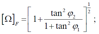
where Φ1 and Φ2 are the geometrical angles for computation defined in Figure 1 and [1-7]. Calculation and programming of these angle ranges is laborious [1-7]. Proof: Complete mathematical development at [1-7]. The extent geometrical elaboration of Omega Factor is shown in Figure 1, presented in previous contributions [1-7].
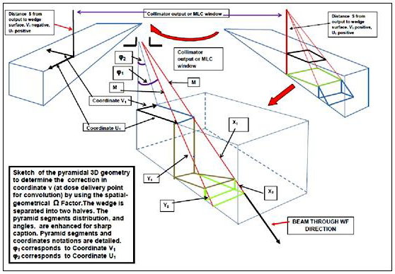
Figure 1: From [refs ], the geometrical exact Omega Factor development. Analytical geometry calculations are rather laborious, [1-7,16-23]. This image is included from previous studies because it shows clearly the Omega Factor geometrical analytic method.
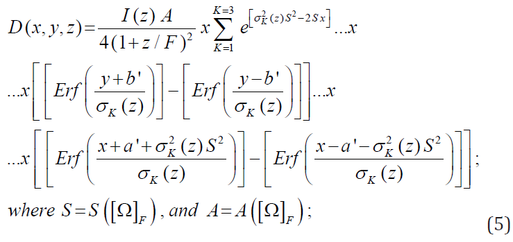
The integral equation for AAA in water with 3D AEF Omega Factor [Ω]F is as follows,
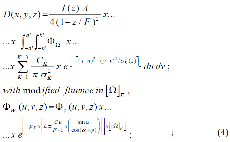
where parameter A is defined for WF [30,31] as,
A = exp[-μW L x (sinα/ (cos (α + φ)) ) ]. Parameters μW (WF material parameter), α (WF angle) and φ (photon-beam divergence angle) are defined at [1-11,20,21]. Development of Equations 1-4 are extensively presented in [1-7]. Finally, the solution, exact, complete, geometrically corrected with Omega Factor, and analytical of this integral equation [Casesnoves, 2015, April, Philadelphia], reads,
Software-Programming Method
The computational method is based on previous software works [1-8,16-23]. This integral equation complete analytical solution, Eq. 5 will be correctly simulated in dosimetry-matrices from 100 x 100 dimensions to 1500 x 1500 dimensions in the following sections and compared with simulations of equations [11-15] of classical AEF [1-7] in AAA model foundations. Further development of Equations 1-5 is extensively presented in [1-7,16-23]. Table 1 shows numerical values implemented for algorithms programming. Table 1 shows the main numerical data for the 3D simulation graphics. The magnitude of Omega factor is about 1.12, for a WF of 15 degrees, and increases with the WF angle till 45° [1-7,16-23]. Note that this apparently small value of Omega Factor becomes propagated by multiplication other constants in formulation and the result is a change of 3D dose delivery magnitude as shown in imaging simulations.
The structure of the program comprises the summatory of every part of Eq. 5. Firstly, these parts of erf functions are set independently one by one. Secondly all of them are summed. Finally, the resulting numerical values are set in the imaging subroutine. These individual erf parts are proven in Table 2. In this study, they can be implemented in Matlab, GNU-Octave, or Freemat. Every system requires an specific modification of the main program to obtain correct simulations. Imaging processing tools and subroutines/options vary in every system. Table 1 shows all the implementation data with corrections from Section 3. Table 2 shows the new model functions [ based in erf function parts for WF] adjusted/improved for setting the new functional software.
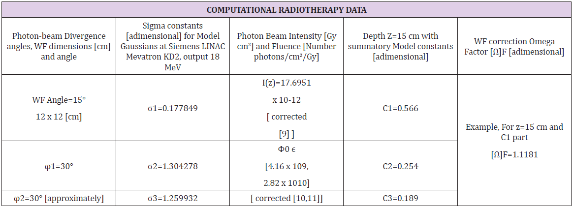
Table 1: AAA model main parameters for 3D Graphical simulations [1-7, 9,10,11, 54, 55].
Note: Data from AAA algorithm foundation in water, [9,54,55], whose numerical computational software used for constants and parameters optimization was Monte Carlo Code EGS-4 and curve fitting MAAFS from CERN (European Union Center for Nuclear Research).
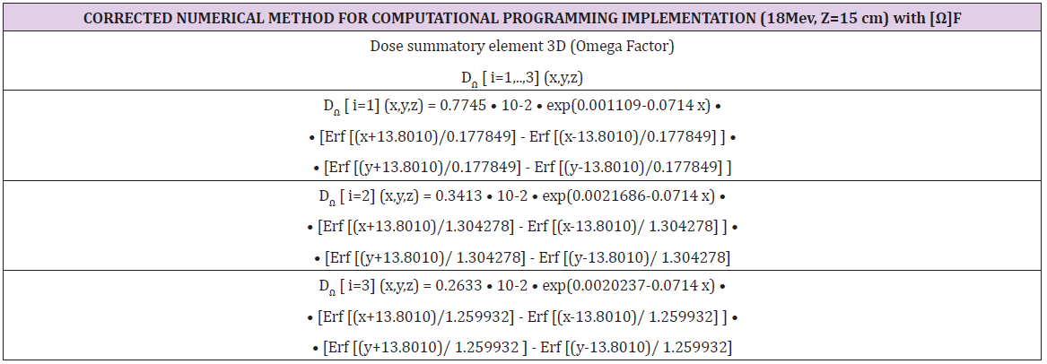
Table 2: Program software parts numerically calculated for Eq. 5 with new corrections [1-7,9,10,11,16-23,54,55]. The data from Section 2, Table 3, was implemented.
I(z) Corrections for AAA Model
After the AAA model foundation, several I(z) important corrections were developed by Ulmer and Harder [9]. Additionally, during 2008-15, a series of geometrical improvements for WF were published [Casesnoves, 1-7,16-23]. These are Omega Factor, Eq. 3, exact path through WF and limit angle for photon-beam through WF [1-7,16-23]. Omega Factor extent calculations and analytical geometry are shown in (Figures 1 & 2). The I(z) corrections were numerically important as I(z) magnitude varied in one magnitude order less. Namely, from 10-11 till 10-12. The corrections were mathematically done, [9], by an exponential-fit in the function I(z) as follows,

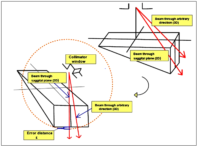
Figure 2: From [1-7], geometrical difference/error-path when using 2D approximation compared to 3D determination. If we take always the AEF approximation of Eq.2, in the sagittal plane, there is an error for less magnitude (blue brackett) in the pathdistance through the wedge. The geometrical exact Omega Factor development. Analytical geometry calculations are rather laborious, [1-7,16-23]. This image is taken from previous publications for paper understanding clarity.
where parameters A, B, a, b, are graphically shown in [9] related to depth-parameter z. Function constants σk(z) were found with no significant numerical differences in photon spectrum [9]. As a result, from this numerical modification, it was found that l(z) changes by up to 0.7 % in the I(z) maximum and up to 5 % at large depth. Table 3 shows these corrections implementation for the 3D simulation study here, with one magnitude order difference [1- 7,9,16-23]. In these simulations fluence [10,11] is set as (particles number/cm2 /Gy). Hence, the numerical modification for fluence factor (FF) magnitude, according to Eqs. 1-5, (Tables 1 & 2), is significant [10,11]. That is, FF ϵ [ 4.16 x 10⁹, 2.82 x 10¹⁰], and it is taken the average. Just remark that the objective of the research is to demonstrate utility and efficacy for clinical/research 3D Graphical Optimization with several systems, not a dose-delivery extremely precise calculation [32-55]. However, acceptable numerical dose delivery data were obtained, (Figures 3-12).
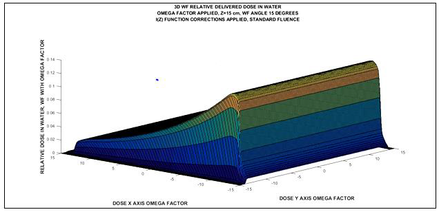
Figure 3: Matlab simulation 3D image for 18Mev photon-beam at 15 cm depth-dose with Omega Factor and I(z) corrected. Fluence magnitude according to Section 3. Matrices for Image Processing have about 103 elements. Imaging Processing Method 1.
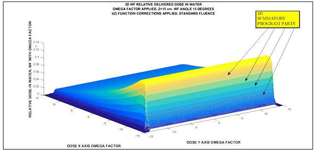
Figure 4: Matlab simulation 3D image for 18Mev photon-beam at 15 cm depth-dose with Omega Factor and I(z) corrected. Fluence magnitude according to Section 3. Matrices for Image Processing have more than 103 elements. Imaging Processing Method 2.
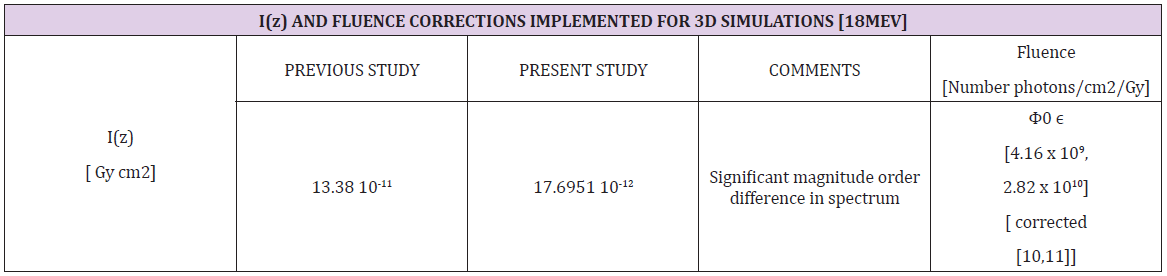
Table 3: I (z) and Fluence numerical corrections [1997,9] related to [54,55,1995-6]. Note the one magnitude order significant difference. This factor has significant influence in photon-dose delivery with/without beam modification devices.
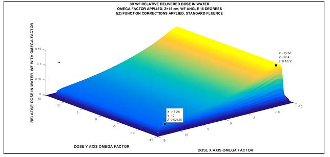
Figure 5: Matlab simulation 3D image for 18Mev photon-beam at 15 cm depth-dose with Omega Factor and I(z) corrected. Numerical data that can be obtained, pictured inset, with image processing method. Relative dose in one-percent, at Z axis, X coordinate at WF, Y coordinate at WF, and program part relative dose. Fluence magnitude according to Section 3. Matrices for Image Processing have about 103 elements. Imaging Processing Method 1.

Figure 6: Matlab simulation 3D image for 18Mev photon-beam at 15 cm depth-dose with Omega Factor and I(z) corrected. Numerical data that can be obtained in simulation comprises four dimensions. Namely, relative dose in one-percent, X coordinate at WF, Y coordinate at WF, and program part relative dose. Fluence magnitude according to Section 3. Matrices for Image Processing have about 103 elements. Imaging Processing Method 1.
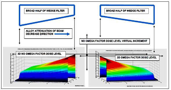
Figure 7: From [1-7], comparative Matlab simulations 3D image for 18Mev photon-beam at 15 cm depth-dose with/without Omega Factor. Matrices for Image Processing have 106 elements. Computational-Graphical simulation-proof of virtual dose error caused by 2D approximated integral equation solution. This simulation is important, because it proves sharply the virtual dose error that is given by the AAA algorithm when using AEF in 2D. That is, the planner system calculates a higher dose compared to the true dose, and this error causes under-dosage on the tumor. On the opposite, 3D planning with Omega Factor results in more precise dose for radiotherapy optimization -with the significant mention that all these calculations and simulations are carried out in water with the foundation AAA model. The simulation is done in the thick part of the wedge, because recent advances have been useful to find a difference in the sign of angle φ1 for the thin half of the WF –this extent analytical-geometry calculation will be explained and simulated in next contributions. The most important objective of this article was to demonstrate the correct approximations and mathematical development together with the computational proof that validates the difference of magnitude between 2D AAA in water and 3D with Omega Factor dosimetry in the same conditions. Note that this picture belongs to former publications without numerical Section 3 corrections.
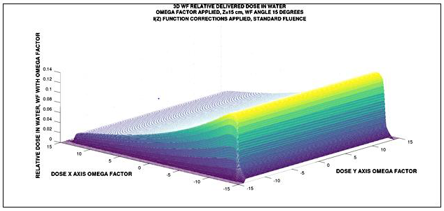
Figure 8: GNU Octave simulation 3D image for 18Mev photon-beam at 15 cm depth-dose with Omega Factor and I(z) corrected. Fluence magnitude according to Section 3. Matrices for Image Processing have more than 103 elements. Imaging Processing Method 1. Image has suitable quality. Running time is longer [ ≈ 20 s ] than Matlab. Software design is very similar.

Figure 9: GNU Octave simulation 3D image for 18Mev photon-beam at 15 cm depth-dose with Omega Factor and I(z) corrected. Fluence magnitude according to Section 3. Matrices for Image Processing have more than 103 elements. Imaging Processing Method 2. Image has suitable quality. Running time is longer [ ≈ 30 s ] than Matlab. With Method 2, the program setting for labels are different.
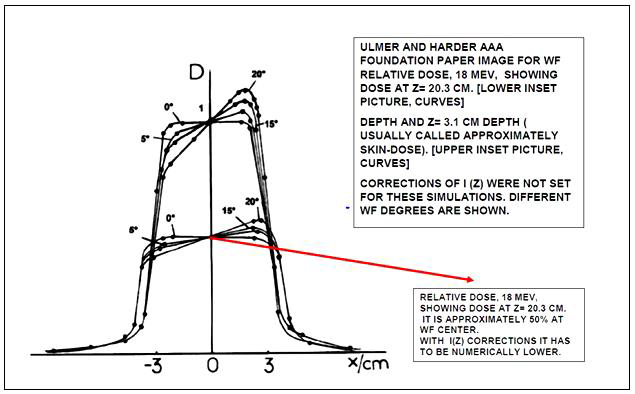
Figure 10: From ref [54], it is sketched a graphical composition from its Figure 5 [Ulmer and Harder 1996]. In this 2D simulation, with non-corrected I (z) function, the dose for WF,18 Mev, at z=20.3 cm (deeper than 15 cm) is around 50% of maximum dose. Numerical 3D Graphical simulations for this study show a dose of about 20% related to maximum dose with I(z) adjustments. In addition, when field size increases, as it is here [ 12 x 12 cm], compared to [-3 , 3], the relative dose generally decreases. Therefore, it is straightforward to guess that approximations of the numerical and imaging software/model are acceptable. Important remark: the very good image/simulation from Ulmer and Harder [54] has a numerical inconsistency at coordinate x=0. That is, the dose at center through WF of different angles cannot be exactly equal. The probable reason is that in the graphical sketch the points at around (-3) and (+3) were matched with curves. That caused quality of dose for different angle WF at x=0 and along D axis.
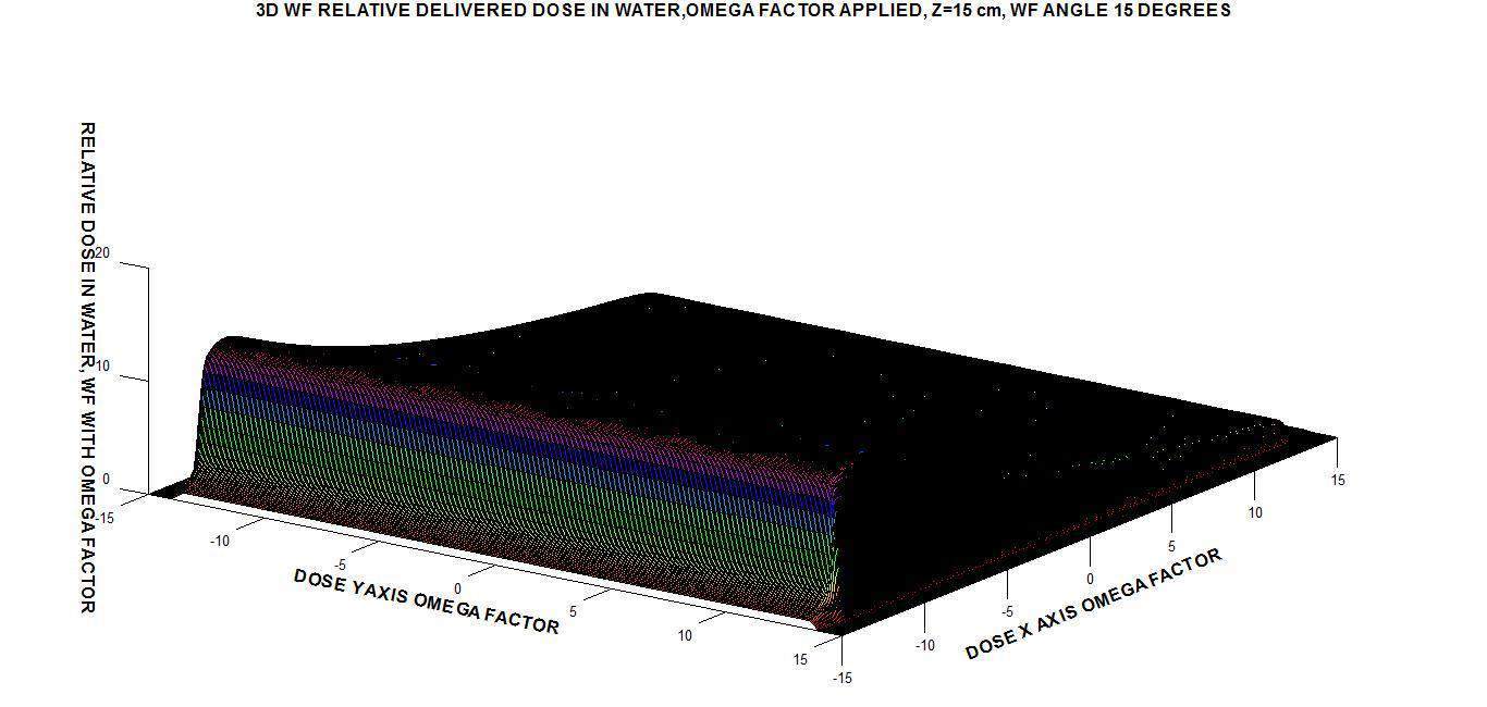
Figure 11: Freemat simulation 3D image for 18Mev photon-beam at 15 cm depth-dose with Omega Factor and I(z) corrected. Fluence magnitude according to Section 3. Matrices for Image Processing have more than 103 elements. Imaging Processing Method 2 [the unique possible in Freemat]. Image is good but slow to obtain, 102 scale factor at Z axis was necessary to get a visible 3D image—that is, expressing dose in percentage. Running time is longer [ ≈ 40 s ] than Matlab and GNU-Octave. With Method 2, the program setting for labels are different. The imaging processing tools are much slower/difficult than Matlab and GNU-Octave. Color and program parts definition are worse than Matlab and GNU-Octave. Relative dose is not higher than 20% maximum dose.
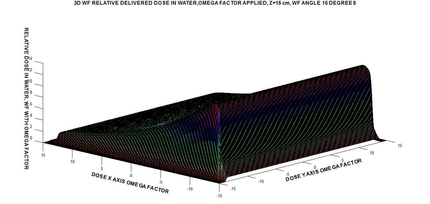
Figure 12: Different view from Fig 11, for showing program parts. Freemat simulation 3D image for 18Mev photon-beam at 15 cm depth-dose with Omega Factor and I(z) corrected. Fluence magnitude according to Section 3. Matrices for Image Processing have more than 103 elements. Imaging Processing Method 2 [the unique possible in Freemat]. Image is good but slow to obtain, 102 scale factor at Z axis was necessary to get a visible 3D image—that is, expressing dose in percentage. Running time is longer [ ≈ 40 s ] than Matlab and GNU-Octave. With Method 2, the program setting for labels are different. The imaging processing tools are much slower/difficult than Matlab and GNU-Octave. Color and program parts definition are worse than Matlab and GNU-Octave.
The model that is implemented in simulations is the original AAA one with Omega Factor [Casesnoves, 2015, 1-7,16-23] WF geometrical correction for water. This model is the base for further developments carried out in foundations [9,54,55]. Besides and later on, superposition-convolution analytical models for proton therapy emerged from this AAA mathematical framework [12]. Among them, tissue inhomogeneities, scatter radiation, or contaminating electrons [9-11]. That is, primary photons and extra-focal photons, scattered in the flattening filter or the beam collimator, flattering filters, jaws, blocks, multi-leaf collimator, and the group of direct dose delivery beam modificators, such as WF, satellite filters, shielding blocks, rectangular satellite filters, etc [14,8-11]. According to all these physical constraints, in practical clinical medical physics treatment planning implementation dose is calculated,

3D MATLAB WF Simulations Results
This section shows the Matlab results for 3D Treatment planning Optimization images and numerical data, (Figures 3-7) with two software methods. (Figure 7) is a comparative sketch to clarify the difference of dose between classical AAA equations and equations modified by Omega Factor [1-7]. The fourth dimension of photon dose is explained in (Figure 6).
3D Gnu-Octave WF Simulations Results
This section shows the GNU-Octave results for 3D Treatment planning Optimization images and numerical data, (Figures 8-10) demonstrates and explains the numerical results acceptable precision. Two software methods are shown. Figure 10 is a comparative sketch with [55] to clarify the small difference of dose between classical AAA WF simulations equations and these 3D imaging simulation results.
3D Freemat WF Simulations Results
This section shows the Freemat results for 3D Treatment planning Optimization images and numerical data, (Figures 11 & 12). For these imaging simulations, Freemat is not as much suitable than Matlab abd GNU-Octave.
Comparative Evaluation
Table 4 shows a comparative assessment for the applied systems. In terms of practical visualization, all systems can be considered acceptable. However, MATLAB makes the best and fast images with data adquisition facilities. The lowest is Freemat, and for this system it is necessary to scale the axes magnitudes to display suitable images. The factor of personal preferences of the programmer for system choice can be considered also. Freemat has exclusively one subroutine imaging option for this 3D simulations.
Clinical Medical Physics Applications
Table 5 shows a resume of principal applications of the results and software methods. Extrapolated applications, e. g. IMRT or IMPT, can also be guessed for future clinical/research radiotherapy developments.
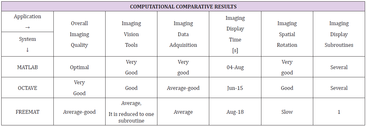
Table 4: Assessment of the selected systems for 3D imaging simulations.
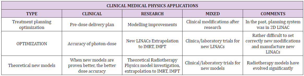
Table 5: Radiotherapy Medical Physics study applications.
Discussion and Conclusion
This study has shown advantages/inconvenients for 3D computational simulations of WF dose delivery with a 18 Mev photon beam at 15cm depth-dose with three computational software-systems. These 3D simulations are corrected in approxitions for several reasons. I(z) important corrections were set in software codes [9]. Fluence has also been modified for better [10,11]. Others are Omega Factor, Eq. 3, exact path through WF and limit angle for photon-beam through WF [1-7,12-19]. In terms of practical visualization, all systems can be considered acceptable. However, Matlab makes the best and fast images with data adquisition facilities. The lowest is Freemat, and for this system it is necessary to scale the axes magnitudes to display suitable images. MATLAB provides with good-quality images, and is considered the best system for these type 3D simulations.
GNU-Octave visualization and 3D plots for photon-dose delivery is good also, while Freemat can be considered a bit more difficult and slower. However, in general, all systems studied are considered acceptable. The selection of any of them depends on computational facilities, programming skills, and image-quality/ vision processing requirements. Tissue inhomogeneities, scatter radiation, or contaminating electrons modifications for 3D graphics have not been applied. The study has proven that there are multiple computational systems to simulate 3D WF conventional photon dose delivery with AAA model—and extrapolation to other models, included IMRT and IMPT. Matlab, GNU-Octave, and Freemat are suitable systems in order of acceptable functionality. High-quality 3D imaging processing methods were proven. 4D image settings constitute an innovation compared to previous contributions [1-7,12-19]. For simulations of dose delivery without beammodification devices, the method is also useful/efficacious.
MATLAB and GNU-Octave shows 2 types of imaging subroutines, while Freemat only one. Medical Physics applications are theoretical and clinical-practical. Theoretical ones involve research in TPO, simulations, implementation in software planning systems, LINAC calibration improvements, photon-dose theoretical-experimental fitting, development/comparison of new optimization methods, and several others. Clinical-practical comprise TPO routine work, clinical medical physics improvements for service functionality, training in delivery simulations, etc. In summary, this imaging radiation therapy WF study shows modern systems for 3D photondose simulation. Imaging processing and computer vision methods are demonstrated. Multiple applications in Clinical and theoretical Medical Physics and Bioengineering emerge from the results.
Scientic Ethics Standards
This contribution is based on Graphical Visualization and Software Optimization methods for radiotherapy modelling improved from previous articles [1-7,12-19]. Graphical- Optimization Methods were created by Francisco Casesnoves in December 2016. The image processing and computer vision tools programs and special software to obtain new dosimetry images positioning, panoramic vision, enhancement of selected WF dose-deposition parts, or imaging tiles optimization was originally developed by author in Matlab, GNU-Octave and Freemat. This advanced article has a few previous paper formulation information, [1-7,12-19], whose inclusion is essential to make the contribution understandable. (Figures 1, 2 & 7) were taken for essential understanding from [1-7,12-19]. This study was carried out, and their contents are done according to the European Union Technology and Science Ethics. Reference, ‘European Textbook on Ethics in Research’. European Commission, Directorate-General for Research. Unit L3. Governance and Ethics. European Research Area. Science and Society. EUR 24452 EN. Also based on The European Code of Conduct for Research Integrity. Revised Edition. ALLEA. 2017. Revised Edition. ALLEA [27,28].
The applications section has some mandatory words from previous contributions. This research was completely done by the author, the software, calculations, images, mathematical propositions and statements, reference citations, and text is original for the author. When a mathematical statement, proposition or theorem is presented, demonstration is always included. The Omega Factor demonstration is not included as it is rather large and can be found at [1-7]. The primary Omega Factor calculations were obtained during MSc Thesis in 1999. The article is exclusively scientific, without any commercial, institutional, academic, political, religious, or economic influence. When anything is taken from a source or previous contribution, it is adequately recognized [29-59].
References
- Casesnoves F (2016) Mathematical Exact 3D Integral Equation Determination for Radiotherapy Wedge Filter Convolution Factor with Algorithms and Numerical Simulations. Journal of Numerical Analysis and Applied Mathematics 1(2): 39-59.
- Casesnoves F (2015) Radiotherapy Conformal Wedge Computational Simulations,Optimization Algorithms, and Exact Limit Angle Approach, International Journal of Scientific Research in Science, Engineering and Technology(IJSRSET) 1(2): 353-362.
- Casesnoves F (2015) "Improvements in Simulations for Radiotherapy Wedge Filter dose and AAA-Convolution Factor Algorithms", International Journal of Scientific Research in Science, Engineering and Technology (IJSRSET).
- Casesnoves F (2011) 'Exact/Approximated Geometrical Determinations of IMRT Photon Pencil-Beam Path Through Alloy Static Wedges in Radiotherapy Using Anisothropic Analytic Algorithm (AAA)’. Peer-reviewed ASME Conference Paper. ASME 2011 International Mechanical Eng Congress. Denver. USA. IMECE2011-65435.
- Casesnoves F (2012) Geometrical Determinations of Limit angle (LA) related to maximum Pencil-Beam Divergence Angle in Radiotherapy Wedges’. Peer-reviewed ASME Conference Paper. ASME 2012 International Mechanical Eng Congress. Houston. USA. IMECE2012-86638.
- Casesnoves F (2013) A Conformal Radiotherapy Wedge Filter Design. Computational and Mathematical Model/Simulation’ Casesnoves, F. Peer-Reviewed Poster IEEE (Institute for Electrical and Electronics Engineers), Northeast Bioengineering Conference. Syracuse New York, USA.
- Casesnoves F (2014) Mathematical and Geometrical Formulation/Analysis for Beam Limit Divergence Angle in Radiotherapy Wedges. Peer-Reviewed International Engineering Article. International Journal of Engineering and Innovative Technology (IJEIT) 7(3).
- Ma C, Lomax T (2013) Proton and Carbon Ion Therapy. CRC Press.
- Ulmer W, Harder D (1995) A triple Gaussian pencil beam model for photon beam treatment planning. Med Phys 5(1): 25-30.
- Ulmer W, Harder D (1996) Applications of a triple Gaussian pencil beam model for photon beam treatment planning. Med Phys 6(2): 68-74.
- Ulmer W, Pyyry J, Kaissl W (2005) A 3D photon superposition/convolution algorithm and its foundation on results of Monte Carlo calculations. Phys Med Biol 50(8): 1767-1790.
- Ulmer WHarder D (1997) Applications of the triple Gaussian Photon Pencil Beam Model to irregular Fields, dynamical Collimators and circular Fields. Phys Med Biol.
- Casesnoves F (2014) Geometrical determinations of IMRT photon pencil-beam path in radiotherapy wedges and limit divergence angle with the Anisotropic Analytic Algorithm (AAA)' Casesnoves, F. Peer-Reviewed scientific paper, both Print and online. International Journal of Cancer Therapy and Oncology 2(3): 02031.
- Casesnoves F (2014) Radiotherapy Conformal Wedge Computational Simulations and Nonlinear Optimization Algorithms’. Casesnoves, F. Peer-reviewed Article, Special Double-Blind Peer-reviewed paper by International Scientific Board with contributed talk. Official Proceedings of Bio- and Medical Informatics and Cybernetics: BMIC 2014 in the context of the 18th Multi-conference on Systemics, Cybernetics and Informatics: WMSCI 2014 July 15 - 18, 2014, Orlando, Florida, USA.
- Casesnoves F (2007) Large-Scale Matlab Optimization Toolbox (MOT) Computing Methods in Radiotherapy Inverse Treatment Planning’. High Performance Computing Meeting. Nottingham University.
- Casesnoves F (2008) A Computational Radiotherapy Optimization Method for Inverse Planning with Static Wedges’. High Performance Computing Conference. Nottingham University.
- Casesnoves F (2015) Radiotherapy Conformal Wedge Computational Simulations, Optimization Algorithms, and Exact Limit Angle Approach ‘. International Journal of Scientific Research in Science, Engineering and Technology. Publication Details.
- Casesnoves F (2015) Radiotherapy Standard/Conformal Wedge IMRT-Beamlet Divergence Angle Limit Exact Method, Mathematical Formulation, and Bioengineering Applications’. International Article-Poster. Published in Proceedings of Conference. 41st Annual Northeast Bioengineering Conference. Rensselaer Polytechnic Institute. Troy, New York USA.
- Casesnoves F (2015) Radiotherapy Standard/Conformal Wedge IMRT-Beamlet Divergence Angle Limit Exact Method, Mathematical Formulation, and Bioengineering Applications’. IEEE (Institute for Electrical and Electronics Engineers), International Article-Poster.
- Casesnoves, F (2015) ‘Radiotherapy Standard/Conformal Wedge IMRT-Beamlet Divergence Angle Limit Exact Method, Mathematical Formulation’. International Conference on Significant Advances in Biomedical Engineering. 252nd OMICS International Conference.
- Casesnoves F (2001) Determination of absorbed doses in common radiodiagnostic explorations. 5th National Meeting of Medical Physics. Madrid, Spain. September 1985. reatment Planning’. Kuopio University. Radiotherapy Department of Kuopio University Hospital and Radiotherapy Physics Group. Finland.
- Casesnoves FA (2013) Conformal Radiotherapy Wedge Filter Design. Computational and Mathematical Model/Simulation’. Peer-Reviewed Poster IEEE (Institute for Electrical and Electronics Engineers), Northeast Bioengineering Conference. Syracuse New York, USA. Presented in the Peer-Reviewed Poster Session on 6th April 2013.
- Vagena E, Stoulos S, Manolopoulou M (1993) GEANT4 Simulations on Medical LINAC operation at 18MV: experimental validation based on activation foils. Radiation Physics and Chemistry.
- Ulmer W, Schaffner B (2011) Foundation of an analytical proton beamlet model for inclusion in a general proton dose calculation system. Radiation PhysicsandChemistry 80: 378-389.
- Sharma SC (2008) Beam Modification Devices in Radiotherapy. Lecture at Radiotherapy Department, PGIMER. India.
- Barrett A Colls (2009) Practical Radiotherapy Planning (4th)., Hodder Arnold.
- (2010) European Textbook on Ethics in Research. European Commission, Directorate-General for Research. Unit L3. Governance and Ethics. European Research Area. Science and Society. EUR 24452.
- ALLEA (2017) The European Code of Conduct for Research Integrity, Revised ed.; ALLEA: Berlin Barndenburg Academy of Sciences.
- Casesnoves F (2014) Die numerische Reuleaux-Methode Rechnerische und dynamische Grundlagen mit Anwendungen (Erster Teil).
- Ulmer W, Harder D (1997) Corrected Tables of the Area Integral I(z) for the Triple Gaussian Pencil Beam Z Med Phys 7: 192-193.
- Haddad K, Anjak O, Yousef B (2014) Neutron and high energy photon fluence estimation in CLINAC using gold activation foils. Reports of practical oncology and radiotherapy 24(1): 41-46.
- Ahnesjö A, Saxner M, A Trepp (1992) A pencil beam model for photon dose calculations. Med Phys 19(2): 263-273.
- Brahme A (2000) Development of Radiation Therapy Optimization. Acta Oncologica 39(5): 2000.
- Bortfeld T, Hong T, Craft D, Carlsson F (2008) Multicriteria Optimization in Intensity-Modulated Radiation Therapy Treatment Planning for Locally Advanced Cancer of the Pancreatic Head. International Journal of Radiation Oncology and Biology Physics 72(4): 1208-1214.
- Brown, Bernardette (2014) Clinician-led improvement in cancer care (CLICC) -testing a multifaceted implementation strategy to increase evidence-based prostate cancer care: phased randomised controlled trial - study protocol’. Implementation Science 9: 64.
- Bortfield T (2006) IMRT: a review and preview. Phys Med Biol 51(13): R363-R379.
- Censor Y, S A Zenios (1997) Parallel Optimization: Theory, Algorithms and Applications'. UOP.
- Censor Y (2005) Mathematical Optimization for the Inverse problem of Intensity-Modulated Radiation Therapy'. Laboratory Report, Department of Mathematics, University of Haifa, Israel.
- Capizzello A, Tsekeris PG, Pakos EE, Papathanasopoulou V, Pitouli EJ (2006) Adjuvant Chemo-Radiotherapy in Patients with Gastric Cancer. Indian Journal of Cancer 43(4): 174-179.
- Tamer Dawod, EM Abdelrazek, Mostafa Elnaggar, Rehab Omar (2014) Dose Validation of Physical Wedged symmetric Fields in Artiste Linear Accelerator. International Journal of Medical Physics, Clinical Engineering and Radiation Oncology 3: 201-209.
- Do, SY, David A, Bush Jerry D Slater (2010) Comorbidity-Adjusted Survival in Early-Stage Lung Cancer Patients Treated with Hypofractioned Proton Therapy. Journal of Oncology.
- Ehrgott M, Burjony, M Radiation Therapy Planning by Multicriteria Optimization. Department of Engineering Science. University of Auckland. New Zealand.
- Ezzel GA (1996) Genetic and geometric optimization of three-dimensional radiation therapy treatment planning. Med Phys 23(3): 293-305.
- (2008) Effective Health Care, Number 13. Comparative Efectiveness of Therapies for Clinically Localized Prostate cancer.
- Silvia C Formenti, Sandra Demaria (2013) Combining Radiotherapy and Cancer Immunotherapy: A Paradigm Shift Silvia C. Formenti, Sandra Demaria. J Natl Cancer Inst 105(4): 256-265.
- Hansen P (1998) Rank-deficient and discrete ill-posed problems: numerical aspects of linear inversion. SIAM monographs on mathematical modelling and computation.
- Hashemiparast SM, Fallahgoul H (2011) Modified Gauss quadrature for ill-posed integral transform. International Journal of Mathematics and Computation.
- Isa N (2014) Evidence based radiation oncology with existing technology. Reports of practical oncology and radiotherapy 19(4): 259-266.
- Johansson KA, Mattsson S, Brahme A, Turesson I (2003) Radiation Therapy Dose Delivery. Acta Oncologica 42(2): 85-91.
- Khanna P, Blais N, Gaudreau PO, Corrales Rodriguez L (2016) Immunotherapy Comes of Age in Lung Cancer. Clinical Lung Cancer 18(1): 13-22.
- Kufer KH, Hamacher HW, Bortfeld T (2005) A multicriteria optimisation approach for inverse radiotherapy planning. University of Kaiserslautern, Germany.
- Kirsch A (1996) An introduction to the Mathematical Theory of Inverse Problems'. Springer Applied Mathematical Sciences.
- Luenberger DG (1989) Linear and Nonlinear Programming (2nd)., Addison-Wesley.
- Moczko JA, Roszak A (2006) Application of Mathematical Modeling in Survival Time Prediction for Females with Advanced Cervical cancer treated Radio-chemotherapy. Computational Methods in science and Technology 12(2): 143-147.
- Numrich, RW (2010) The computational energy spectrum of a program as it executes. Journal of Supercomputing.
- Joseph Ragaz, Ivo A Olivotto, John J Spinelli, Norman Phillips, Stewart M Jackson, et al. (2005) Locoregional Radiation Therapy in Patients with High-risk Breast Cancer Receiving Adjuvant Chemotherapy: 20-Year Results of the Columbia Randomized Trial. Journal of National Cancer Institute 97(2): 116-126.
- Steuer R (1986) Multiple Criteria Optimization: Theory, Computation and Application.
- Spirou SV, Chui CS (1998) A gradient inverse planning algorithm with dose-volume constraints. Med Phys 25(3): 321-323.
- Sievinen J, Waldemar U, Kaissl W AAA Photon Dose Calculation Model in Eclipse™. Varian Medical Systems Report Rad.

Radiotherapy Wedge Filter AAA Model 18 Mev- Dose Delivery 3D Simulations with Several Software Systems for Medical Physics Applications
ABSTRACT
Along a series of previous contributions for Anisotropic Analytic Model (AAA) improvements, several exact/approximated formulations/corrections for wedge filters (WF) photon-dose delivery were presented. Namely, exact photon-beam pathlength through wedge, exact beam limit divergence angle algorithm, and dose delivery correction Omega Factor for wedge filters. Based on all these algorithms/software, a number of 3D comparative-optional simulations with several software systems are developed for AAA model 18 Mev photon-beam. Explicitly, Matlab, Freemat and GNU Octave systems. In these new simulations algorithm, the corrected AAA photon beam intensity I(z) magnitude modification, fluence and geometrical Omega Factor are implemented. Results comprise a series of optimized 3D graphics with numerical data for AAA photon-dose delivery modified by WF. Computational findings show 3D charts for all these programming systems. New results with 4D Interior Optimization imagingdevelopment are presented with software. Resulting consequences for comparative evaluation of the programming methods with all systems are explained. Radiotherapy Medical Physics applications for wedge filter photon-dose calculations emerge from all the numerical and graphical outcomes. Extrapolated clinical radiotherapy applications are obtained from 2D/3D graphical simulation series.
Keywords: Radiation Dose; Attenuation Exponential Factor (AEF); Simulations; Nonlinear Optimization, Matrix Algebra; Spherical-Spatial Analytical Geometry; Series Approximations; Multi-Leaf Collimator (MLC); Wedge Filter (WF); Conformal Wedge Filter; Anisotropic Analytic Model AAA; Intensity Modulated Radiotherapy (IMRT); Intense Modulated Protontherapy (IMPT); Fluence Factor (FF); Treatment Planning Optimization (TPO)