info@biomedres.us
+1 (502) 904-2126
One Westbrook Corporate Center, Suite 300, Westchester, IL 60154, USA
Site Map

Received: December 01, 2017; Published: December 11, 2017
Corresponding author: Gökmen Akkaya, Department of Cardiovascular Surgery, Ege University School of Medicine, 35040 Bornova/Izmir, Turkey
DOI: 10.26717/BJSTR.2017.01.000577
Objective: Solitary fibrous tumors are mostly benign tumors that originating from submesenchymal cells of parietal or, more commonly, visceral pleura.
Case: In that case, we have hospitalized a male patient, who was attended to the emergency service with massive bleeding, with a giant (15 x 20cm) mass located on his nape. The operation was planned as a collaboration of plastic and cardiovascular surgery teams. After a successful surgical excision histopathologically, the result was solitary fibrous tumor.
Discussion: Our approach resulted in a satisfying operative result without harmful complications in such a difficult case. In our opinion, surgical approach should always be thought as an option regardless of size in case of solitary tumors.
Key words: Solitary Fibrous Tumor; Vascular Surgery; Nape
Solitary fibrous tumors are mostly benign tumors that originating from submesenchymal cells of parietal or, more commonly, visceral pleura. They contain both normal and abnormal vascular structures. Usually, they become symptomatic due to ulcerations, bleeding and compression of surrounding tissues. We have hospitalized a male patient, who was attended to the emergency service with massive bleeding, with a giant (15 x 20cm). Solitary fibrous tumor located on his nape. The case was decided “not suitable for interventional embolization” so it was widely excised by plastic and cardiovascular surgical teams. The tumor was removed by surgery. Its weight was 1788gr. This is a rare clinical pathology, which has not been published in the English literature before.
An 80 years old male patient had a history of CABG and low EF (25%) and was suffering a progressively growing, discoloring mass on his nape since 5. He was followed in a rural hospital for 4 years. He had been forwarded to an advanced center because of the magnitude of the tumor and cardiac condition of the patient. He had major bleeding requiring transfusion one year ago. He was hospitalized in our intensive care unit (ICU) due to massive bleeding. He had a big (15 to 20cm) pulsatile mass on his nape, which was oozing bloody and requiring compression (Figure 1). Vascular supply of the mass was visualized by computerized tomography neuroangiography. Both subclavian arteries (left sided dominancy) were supplying the ulcerative tumor. Due to large diameter supplying arteries and excising blood flow, interventional embolization was unsuccessful. Surgical excision and bleeding control were planned. Vital stabilization was maintained with transfusion of 14 bags of packed red cells and freshly frozen plasma. He had received diuretics and positive isotropic treatment (moderate dose of dopamine -5mcg/ kg/m-) for heart failure and monitorzed in the ICU. The operation was planned as a collaboration of plastic and cardiovascular surgery teams. Voluntary informed consent from the patient received with the teams. The procedure was initiated under general anesthesia and on prone position.
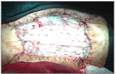
The mass was circumferentially sutured with continued 1/0 vicryl. After skin incision, vascular connections were dissected. It was widely excised. All vascular structures, connected to subclavian and vertebral arteries, were controlled with either electrocautery or 2/0 silk sutures. After safe controlling of the bleeding sources, excision of the mass was concluded (Figure 2) and the 15 cm to 20cm excision material was sent for pathological examination (Figure 3). Plastic surgeons had prepared two fascia-cutaneous flaps from the thigh of the patient to repair the margins of the skin defect and central portion of the defect was cloded with a split thickness skin graft (STDG), which was obtained from his right thigh. Operation was terminated with “tie-over” dressing of the wound (Figure 4). Histopathologically, the result was Solitary fibrous tumor. (cd34, focal+, s100-, desmin-, cd117-, ema-, ki67, bcl2+, keratin-, caldesmon-, cd99-, aktinfokal+). Solitary fibrous tumor of the pleura was rare neoplasms andaccount for less than 4% of all pleural tumors. That originates from mesenchymal cells of the submesothelial tissue of the pleura. He was monitored in the ICU for two days and after an uneventful period of care, transferred to the ward.
Figure 1: Preoperative mass image.
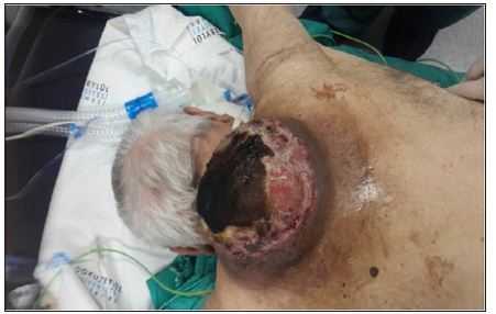
Figure 2: Excised material.
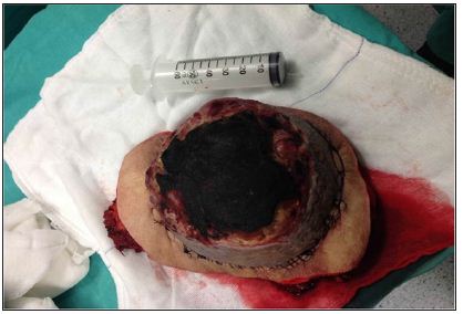
Figure 3: Postoperative tissue defect.
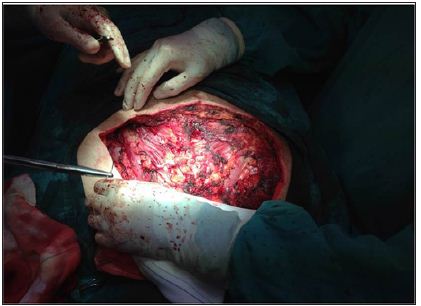
Figure 4: Tissue covered the final image.
Solitary fibrous tumors are mostly benign vascular tumors that characterized with spontaneous regression in 80% of cases [1-3]. Solitary fibrous tumor is a commonly benign vascular proliferative lesion that can be present at any age [2-4]. Lesions usually expand to the surrounding tissue and become ulcerative in several years. Diagnosis of the tumor is based on physical examination [5]. They have very frequent slow secondary regression. There are several therapeutically modalities such as injection of sclerosing solutions and steroids, laser therapy, radical surgery, radiotherapy and embolization. We report an 80 year old case with a giant cutaneous solitary fibrous tumor of 15 x 20cm in diameter, diagnosed 5 years ago, with massive bleeding, requiring transfusions. Physical examination did not show any other cutaneous vascular malformations. He was hospitalized for infection and bleeding of the giant tumor 6 months ago as a cutaneous hemangioma. He had not been intervened because of complexity of the tumor and his cardiac status. We discuss here the diagnostic, therapeutic and operational aspects of this pathology and review the literature. Solitary fibrous tumors have to be carefully followed for evaluation. Bleeding and compression of surrounding tissues may result in catastrophic events. Best results can be obtained with surgery. In this case, we have not observed any residual lesions. Circular excision and purse-string suturing technique were safe to control bleeding. Large scar after excision can be easily repaired by plastic and reconstructive surgery. Our approach resulted in a satisfying operative result without harmful complications in such a difficult case. As a result,surgical approach should always be thought as an option regardless of size in case of solitary tumors.


