info@biomedres.us
+1 (502) 904-2126
One Westbrook Corporate Center, Suite 300, Westchester, IL 60154, USA
Site Map

Received: August 09, 2017; Published: August 30, 2017
Corresponding author: Chateen I Ali Pambuk, College of Dentistrym, University of Tikrit, Iraq
DOI: 10.26717/BJSTR.2017.01.000312
This study aims to measure the levels of fat in women before and after menopause, which is considered as an indicator to increase the likelihood of cardiovascular disease. This study included 210 Iraqi women before menopause, which ranged in age between (27-45), and 200 of postmenopausal women were between the ages of (51-67). Blood samples were collected for cholesterol measurement, triglycerides and high protein density and low protein density and low-density lipoprotein too. The results showed no significant differences in cholesterol TC level as the results of the study showed that there are statistically significant differences for triglycerides (TG) which reported results significantly increased (P <0.05) in postmenopausal women when compared to pre menopausal women in which the results were (145.97±39.64) (100.87 ±32.11) mg / dL, respectively. As for (HDL-C) there was a significant decrease (P <0.05) in postmenopausal women (34.41 ± 10.92) when compared with pre menopausal women (43.01 ± 14.29), as well as the case for (LDL-C) in which there was a statistically significant increase (P <0.05) with reported results (108.59±21.20) in postmenopausal women and (77.11 ± 30.36) in women before menopause. Regarding (VLDL-C) in postmenopausal women, the results recorded a significant increase (P <0.5) in terms of its percentage (32.93±8.44) when compared with women before menopause (21.31 ± 7.12). We conclude in this study, the presence of statistically significant changes in the proportions of fat, which is considered a risk factor for blood vessel and heart disease in Iraqi’s women.
Keywords: Serum Lipid Profile; Premenopausal; Postmenopausal Women
Menopause is the cessation of menoustral cycle in women. It represents a transitional phase in the natural biological woman’s age, a natural event that marks the end of the reproductive years of women in terms of the menstrual cycle permanently stopped and the resulting stop ovarian function in females [1]. Most of the women at this stage suffer from a variety of symptoms due to hormonal changes and these symptoms are possible to be severe and frequent, some women have more severe symptoms and others do not suffer from any symptoms at all [2]. This age can be divided into: the period before menopause (Pre menopause) representing any reproductive period before menopause they represent years before the last menstrual period [3]. And to the period after menopause (Post Menopause), is the period of a woman’s life that comes after the last menstrual cycle, i.e. when the ovaries completely stop working, regardless of the reasons for the drop naturally or because of disease [4].
The increase in the proportion of lipid in the body is a major cause of heart and blood vessels diseases and is the most common, where it causes deaths in females after menopause [5]. Imbalance and Dyslipidemia in fat state increases the risk of cardiovascular disease. Cholesterol is an organic molecule found in food and can be slowly absorbed from the gastrointestinal tract. Is the compound found in all cells of the body and has a vital importance and greatly plays a major role in membranes of real nucleus eukaryotes which constitute the majority of the fat in the body [6]. As LDL which is the bulk of the cholesterol in blood and represents bad cholesterol carried by low-density lipoproteins LDL and represents the spherical particles contain cholesterol ester works to transfer cholesterol from the liver to the tissues. The HDL is working on the transfer of low-density lipoprotein LDL from the blood to the liver to get rid of it, prevents the deposition of cholesterol in the walls of the arteries [7].
Triglycerides are considered as a great source for energy storage. It will give rise to an organic compound consisting link glycerol with three molecules of fatty acid. Triglyceride moving through the blood and stored in the fat cells. Triglyceride posses an important functions in the body which provides the body with vigor and energy, several studies demonstrated a relationship between triglycerides and LDL and heart disease [8]. So far, most studies suggest the effect of triglyceride increase on the risk of cardiovascular disease [9]. The present study is undertaken to measure levels of serum lipids in postmenopausal Iraqi women and compared with pre-menopause in Sulaymania city.
This study was conducted in the city of Sulaymania on healthy women before and after menopause for the period from January (2015) until June (2015). Samples were selected randomly from healthy women visitors or patient’s escort in private clinics, and attenders to private and general hospitals and their companions. The he collection of samples taken from different parts of the city of Sulaymania. Blood samples underwent for analytical laboratory blood tests in the in a specialized laboratory in Sulaymania general hospital. A total of (210) women samples were chosen in the age group (27-45 years) representing a class of women before menopause, also (200) postmenopausal women Samples were selected in the age group ranging from 51 to 67 years.
Information recorded on paper questionnaire (Questionnaire sheet) and thoroughly for each sample selected. Samples Chosen depending on the selection and survey standards, information was collected on social and demographic characteristics (Sociodemographic Characteristics) and age (Age), weight, height and lifestyle (Life Style), and menopausal status. A written informed consent was obtained from all women under study. Those who suffer from disorders in fat and those who suffer from diabetes disease and high blood pressure, kidney pressure, cardiovascular and inflammation of acute and various liver diseases and chronic diseases, have all been excluded from the study. Lipid measurement Cholesterol, LDL, HDL and triglycerides were measured by the same methodology by using a special device called Cobas C111, and according to the manufacturer’s directions for several work (Kit’s) special for each type of lipid , and the details were followed as follows: Blood was withdrawn hours after fasting (10-14 hours) for both groups before and after menopausal women . blood in tubes were left at room temperature for (15) minutes to form a clot and then was run in the centrifuge for a period of [10] minutes at 5000 rpm in order to obtain the serum . About 850 μl of serum sample was put and placed in a small tube Cuvette, and put in the allocated place in the machine (Cobas C111) and adjusted for the following analyzes for TG - LDL - HDL - CHOL, then the results were recorded.
CHOL2 (Cholesterol Gen2) was used for the quantitative measurement of cholesterol in the serum of individuals under study by using (Hitach Cobas C111) (Rochel). The basic principle of this measurement is the (Enzymatic colorimetric methods) in which the hydrolysis of esters of cholesterol by cholesterol esterase were done forming cholesterol and fatty acids, and then the oxidation of cholesterol by cholesterol oxidase to Cholest-4-en -3- one and hydrogen peroxide (hydrogen peroxide H2O) formed affect the pairing oxidative phenol and (4-amino anti pyrine) to form a red stain (Quinone - amino). The density of color to the formula formed directly proportioned to the concentration of cholesterol. This was determined by measuring the increase in absorbance by the following equations:

Cholesterol esters + O2Cholest -4- en – 3 one + H2O

TRIGIL been used of the quantifiable (TG) in the serum of individuals under study using Rochel / HitachCobas C111 device. The principle of quantitative analysis of (Triglycerid) in this method is the use of an enzyme Lipoprotein Lipase, used in microbiology for full rapid analysis of (Triglycerids) to Alklserol Glycerol, followed by the oxidation process of Glycerol to form (Dihydroxy acetone phosphate) and Hydrogen Peroxide H2O2, though H2O2 formed in this process reacts with (4-amino phenazone) and (4-cholrophenol) and the presence of peroxidase enzyme (peroxidase) to form a composite red color. The intensity of the color of dye formed inversely proportional and directly with the concentration of triglycerides (TG) and is determined by measuring the increase in absorbance and by the following equations:
Enzymatic Colorimetric Test:


Glycerol -3- phosphate + O2dihydroxy acetone phosphate+H2O2
The use of HDL-C3 (HDL - Cholesterol) is so quantifiable for HDL in the serum of individuals under study using Rochel / HitachCobas C111 device. The basic principle of this measurement is the (Enzymatic colorimetric methods) in which in the presence of magnesium ions, the Dextran Sulfate be complexes soluble in water with the (HDL) and the (VLDL) are appreciated enzymatically by an enzyme (Cholesterol esterase) and (Cholesterol oxidase) associated with (PEG). The Cholesterol esters are quantitatively broken down by enzyme (Cholesterol esterase) to form the free acids and cholesterol which in the presence of oxygen cholesterol oxidation occur by the enzyme (Choli Oxidase) to form hydrogen peroxide cholestinon:

Cholistenone+H2O2
In the presence of an enzyme POD Peroiadse the Hydrogen Peroxide formed by the interaction with 4- amino - antipyrine and HSDA to form a blue dye, the color intensity of the dye formed is proportional to the concentration (HDL-C).
LDL-C kit was used for quantitative measures of LDL-C in the serum of individuals under study using Rochel / HitachCobas device C111. The basic principle of this measurement is an Homogeneous enzymatic colorimetric assay. Where cholesterol esters disintegrated quantitatively to form free cholesterol and fatty acids, so the presence of the enzyme (Cholesterol esterase)

Then in the presence of oxygen the enzyme cholesterol oxidase oxidize LDL-cholesterol to cholestinon and H2O2 Hydrogen peroxid

And in the presence of an enzyme peroxide (Peroxidase (POD)) the formed H2O2 in the reaction of (4-amino - anti pyrine) and HSDA, to form a blue dye, the density of the color of the dye formed direct proportion to the concentration of (LDL-C), which is determined through measurement the increase in absorption.
Evaluation of VLDL was calculated according to the following equation

The statistical analysis of the data was performed using statistical analysis program (MINI TAB) according to T_test. The arithmetic mean and standard deviation have been obtained. Use P_value as the level of statistical significance at the level of (p <0.05) and significantly high at the level of (P <0.01).
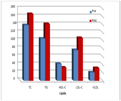
Figure 1: the lipid levels (lipid profile) in the serum of pre and postmenopausal women.
The statistical analysis of the results showed As shown in Table 1 and Figure 1 that the lipid levels were generally higher in postmenopausal women when compared with women before menopause, where it appeared that there was a significant increase, but not significant (P> 0.05) for total cholesterol (TC) in postmenopausal women when compared with a pre-menopausal women, where the proportion of the study recorded (TC) (166.89 ± 43.05) (138.52 ± 38.22) mg / dL, respectively. The results of the present study showed that there are a statistically significant differences for triglycerides (TG), the results reported a significantly increased (P <0.05) in postmenopausal women when compared to women before menopause, where the results were (145.97 ± 39.64) (100.87 ±32.11) respectively.
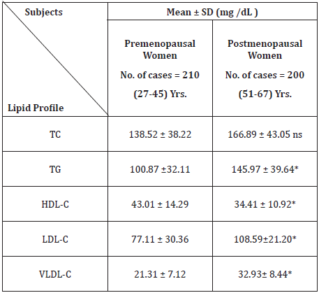
Table 1: The lipid levels (Lipid profile) in the serum of women before and after menopause.
TC: Total cholesterol, TG: Triglyceride, HDL-C: High density lipoprotein - Cholesterol, LDL-C: Low density lipoprotein – Cholesterol, VLDL-C: Very High density lipoprotein - Choesterol
**Highly significant p≤ 0, 01
*Significant p≤ 0.05
ns: Non significant p> 0.05
As for (HDL-C) there was a significant decrease (P <0.05) in postmenopausal women (34.41 ± 10.92) when compared with women before menopause (43.01 ± 14.29), as well as the case for (LDL-C) where there was a statistically significant increase (P <0.05) which reported s (108.59 ± 21.20) in postmenopausal women and (77.11 ± 30.36) in women before menopause. Concerning (VLDL-C) in women after menopause, the results recorded a significant increase (P <0.5) in terms of its percentage (32.93± 8.44) when compared with women before menopause (21.31 ± 7.12). In this respect, pre menopausal group were divided into two age subgroups, from the total samples collected 210 samples, which were between the ages of (27-45) years to, the first set of subgroup was (108) samples ranged in age from (27- 34 years), while the second sub-group was (102) sample aged between (35-45) years, the results shows, and as shown in (Figure 2) the presence of slight increases but statistically was not significant (P> 0.05). The level of TC, TG, HDL-C, LDL-C, VLDL-C in the second age group in women before menopause, and that between the ages of (35-45) years,were (149.17 ± 47.12), (113.32 ± 43.07) , (42.92 ± 18.95), (85.12 ± 30.62), (21.46 ± 8.88) mg / dL when compared with the sub-group for the first of the women before menopause, where the level of the lipids were (130.61 ± 34.19), (95.44 ± 25.76), (46.47 ± 17.96), (66.28 ± 27.39), (19.66 ± 4.88), respectively.
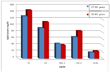
Figure 2: The effect of age groups upon the serum lipid profile in pre menopause.
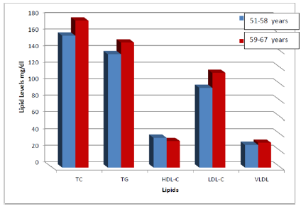
Figure 3: The effect of age groups on lipid levels (lipid profile) in post menopause.
For women after menopause with 200 samples, the women ranged in ages (51-67 years) and as shown in (Figure 3). Postmenopausal women were divided into two subgroups, where the number of the first sub-group (133) sample aged between (51-58) and second sub-group (67) sample aged between (59-67) years. The results in general showed between sub age groups a slight increase in the level of lipids but they were not significant (P> 0.05). Between the two groups the level of each of (TC, TG, HDL-C, LDL-C, VLDL-C) in the second age group after menopause, and that was ages ( 59-67 years) (175.44 ± 47.19), (151.24 ± 35.77), (33.21 ± 9.13), (116.08 ± 26.95), (31.13 ± 7.37) when compared with the first group for age, which ranged in age (51-59 years) the lipid levels (159.19 ± 40.01), (137.27 ± 33.78), (37.04 ± 12.48), (95.29 ± 20.26), (28.35 ± 6.49), respectively. The results of the current study, which was obtained is in accordance with the many analytical data in the world, that these results are compatible with Swapnail et.al [10], where he pointed to a significant increase in the average values of TG, LDL-C, VLDL-C, and recorded the results as well as low levels of HDL-C in postmenopausal women when compared with women before menopause, but the results of the current study contradict this paper [10] in cholesterol TC which showed a results in the proportions of a significant TC ratios (P <0.05), while the results of our study had cholesterol no significant levels (P> 0.05) between the women before and after menopause in general or between the age groups.
The results obtained in this study regarding cholesterol levels agrees with several research articles. The results agreed with the research conducted by Igweh [11] which has recorded similar data values exhibited by the current study, especially in terms of the proportion of the TC where it was fully compatible with the current study, scoring non significant increased levels (P> 0.05) as the results of our study also coincided with the research presented by Usoro et.al [12]. It is worth mentioning that there are differences in the levels of fat that is obtained and recorded in different individuals on the basis of race, age, sex, body mass (obesity), exercise, smoking, alcohol, diet and many diseases such as diabetes, hypertension and chronic liver and kidney disease pressure [13], however, we tried in the current study excluded overlapping variables that increase the statistical error, and after excluding these factors possibly we have taken results obtained scientifically criterion correct natural increase, but the current study data values contrary to numerous studies in different regions of the world with respect to the levels of fat in postmenopausal women.
In a cross-sectional study conducted by Kanwar et. al [14] the differences of data rate and standard deviation of TG values , were not significant in postmenopausal women when compared with women before menopause, as well as levels of VLDL-C, this study reported a significant decrease (P <0.05) in women after menopause when compared with women before menopause, but the study concluded that there were significant differences, which reported a significant decrease in HDL-C and a significant increase in LDL-C and non- significant differences in TC and this agrees with what we got from the results in this study. In another study conducted by Otolorin [15] the results were compatible with the present study in which there was no significant change in the TC cholesterol levels when compared to postmenopausal women with women before menopause. Furthermore, Yassin et.al [16] recorded results which were consistent with the results of the current study for the level of HDL, LDL has earned a morale levels and, on the other hand his results contrasted the findings of the results of our study in each of the TG and TC. The results of our study agreed with Swarna [17] in the levels of each of the TC, HDL, TG, VLDL except LDL level was not significant. This is a contradiction with what we got in our study. In studies conducted in some developed countries centers demonstrated a highest value regarding the level of cholesterol TC so that it reached the boundary of significance which scored by Grundy [18] and this contradicts what we found in the present study, where the proportions of the TC in our study were not significant.
It is worth mentioning in determining the risk of cardiovascular disease, the value of the absolute cholesterol is not the most important factor, but abnormal levels of various concentrations of patterns of cholesterol HDL-c, LDL-C, TG, and comes as a result of ethnic differences and genetic patterns included by other factors intertwined with , the values of this study fit exclusively with women in the province of Kirkuk, and all the differences statistically with similar ethnic and genetic patterns and counterparts in parts of the world, it is worth mentioning as it is estimated that increasing the HDL-C by [1] mg / dL, there is a decline (3%) the risk of coronary artery disease and a decrease of (4.7%), the risk of death from cardiovascular disease [19]. In the current study has been the loss of this protective effect. In our study the percentage of dropped HDL_C increased level of LDL_D this is a large and important indicator of the development of atherosclerosis as it is noted by several studies to this [20], there is no doubt in this study that the changes that occur in the level of fat in the blood after menopause are unhealthy changes and harmful to the health of the heart and blood vessels is generally believed that the postmenopausal symptoms is less in our women in Caucasus and Western counterparts and this is something that also affects different variables in a woman’s body, and this may not be true because many of the global research data refers to other factors, many variables may affect this, including the psychological, physical factor, biochemical , hormonal and motor factors [21]. However, this is may be in aspects similar to these researches or in some be different and the problem may be perhaps is in the economic and social level and extreme climatic and bad conditions, which cast a shadow over many variables in our women and perhaps different to what is found in women in the rest of the world. Furthermore, In this respect, in our previous study we concluded that there is approximately 3 fold significant increase in hs-CRP levels in post menopausal Iraqi’s women, suggesting more burden in Inflammatory process with increase lipid levels and may be Coronary artery diseases CADs [22].
Moreover, the rise in the levels of harmful (LDL-C) which coincides with the decline in the (HDL-C), which is a protective factor against heart disease, in the presence or absence of (VLDL-C), is one of the outstanding relations perhaps for menopause in our society and this independent risk factor itself to cause cardiovascular disease in our society. There is much debate as to whether menopause itself affects the level of fat, and the reasons that explain the differences in fat may be income, household size, and age at menarche, duration of the cycle and regularly or irregularly duration of cycle. As for the differences between the age groups there was an observable but no significant increase in lipid levels and this is in parallel with what other study found [5]. Many studies have shown the beneficial effects of alternative hormone treatment (Hormone replacement - Therapy (HRT) on the lipid levels in the serum of postmenopausal women, however, controversy exists that these variables do eventually lead to a lower risk of heart disease and blood vessels, so a numerous observational studies were conducted over years have emphasized on the hormone replacement therapy beneficial effects (HRT) in the prevention of cardiovascular disease in postmenopausal women [23], and we must point out that these women in Kirkuk, as the results have shown in this study, suffer from an increase in the levels of harmful fat in the blood, which is the basic cause of cardiovascular disease factors and therefore it is very important to keep them under medical eyes in this field and to correct the imbalance in the level of fat, because the using of alternative hormone therapy (HRT) as well as fat lowering drugs is still controversial, so it is very important to the community, institutions and members of the health community awareness in this area to correct paths and eating habits, exercise and the application of preventive measures for a healthy and enjoyable life.
This research was supported by research grants of post graduate studies from The Ministry of higher education and scientific Research of Iraqi Government. Many thanks for all those assist us in the course of our study.


